oh my goodness...this is gold... you are an excellent teacher...very concise...very easy to follow... First year radiology resident here, total rookie at Ultrasound
How about patient has ascites and youre having trouble filling in the vessels ? At times its difficult for me to tell if its thrombosed or not. I drop the PRF still no filling then I increase over color gain and fills in but aliasing appears. I then would increase PRF to get rid of aliasing but then lumen becomes not fully filled 🤦🏻♀️. My question is how can I convince the radiologist I did my absolute best in optimizing the image? Do you have any tips when you run into these kind of difficult cases? Thank you in advance
Love your videos Henry. Thank you so much. Are you able to make a video explaining liver transplant and complications? Would love to see that, thanks again!!!
thank you very much, Henry I like all your videos. you are source no.1 for me may God bless you for all your hard-working..keep on you are helping us a lot.


oh my goodness...this is gold... you are an excellent teacher...very concise...very easy to follow... First year radiology resident here, total rookie at Ultrasound
Thank you so much.. I never learnd sonographic liver segment . İts hard to me . Thank you for beautiful explain🙏🏻🙏🏻
You’re very welcome it took me a while too!!
Is the SMV waveform supposed to be above eor below the baseline? I keep getting contradictory responses.
It should be hepatopetal or towards the liver, so if you get the SMV in a sagittal plane then it should be above baseline
How about patient has ascites and youre having trouble filling in the vessels ? At times its difficult for me to tell if its thrombosed or not. I drop the PRF still no filling then I increase over color gain and fills in but aliasing appears. I then would increase PRF to get rid of aliasing but then lumen becomes not fully filled 🤦🏻♀️. My question is how can I convince the radiologist I did my absolute best in optimizing the image? Do you have any tips when you run into these kind of difficult cases? Thank you in advance
I love your videos. Awesome information for years to come. Students, new grads, and experienced sonographers alike can all learn from you! :)
Thank you!!
Awesome videos Henry 📹 👌 👏 👍 🙌
Love your videos Henry. Thank you so much. Are you able to make a video explaining liver transplant and complications? Would love to see that, thanks again!!!
thank you very much, Henry I like all your videos. you are source no.1 for me may God bless you for all your hard-working..keep on you are helping us a lot.
Thank you, so much. This, and all your videos, are outstanding.
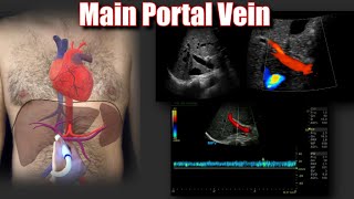
at which part of the portal vein do we measure its diameter ?
At the porta hepatis (entrance to the liver)
Thank you so much for your videos!! I love the tutorials and your funny videos, too! You get helped me do my first Liver Doppler on my own!
Thank you
please can I communicate with you Um Muhammad
Do the portal veins carry oxygenated or deoxygenated blood to the liver?
You’re an incredible teacher - so inspiring. Please never stop, you’re so appreciated!!
6:07 can it be considered a doppler artefact?
Good video
Thanks alot really helpful
Thanks for the detailed insight of hepatic doppler.
Can you show SMV??
Fantastic
Thank u for sharing
Thanks a lot !
Thank you
Thank you!!
Excellent demo
Thanks