The best channel, i did this morning the DVT Poc US i found all the images they are all very good and so nice, was so proud of my self, thank you, tomorrow will try the gall bladder and portal triade
I work in the ER so don't have the luxury of patients fasting before coming in so I get why you are asking this question. I teach my residents (they are learning POCUS) to do the study the same way every time - same sequence of image capture, measurements, etc. This way they are less likely to miss a finding or miss capture of an image. This includes the GB wall measurement. With that said I know many radiology departments that don't measure it unless visually it looks thickened. I personally think the measurement has little value unless the gallbladder is dilated somewhat (arbitrarily I think it should have a diameter of greater than 2 cm before I worry too much about the anterior gb wall -- this is an opinion though.)
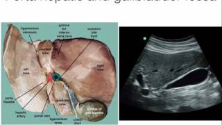
Awsome video, Thanks for share it!... I have Just one question, Wich probe position do you recomend its the apropriate to watch the common bile duct in its transversal aspect and the portal vein in its transversal aspect as well? (The 6th image in the summary chart)


You are definitely the best POCUS UA-camr ever I should learn from.
The best channel, i did this morning the DVT Poc US i found all the images they are all very good and so nice, was so proud of my self, thank you, tomorrow will try the gall bladder and portal triade
Highest regards to you tutor. You are not showing off, you are teaching. God bless you!
As a new Radiology Resident in training, this was such an excellent video! Thank you very much :)
Every time I watch it, it's a good reminder
Amazing video. I’m a first year DMS student and your video and the way your explained it was so helpful. TY!
Thank you, sir. Best video I've seen on the subject hands down.
Definitely the best video on hepatobiliary ultrasound. Thanks alot sir
12:30 thank you for explaining! The whole time I was wondering in which direction the probe was directed!
Excellent teaching Dr Marx! Thank you👍
Thanks sir keep sharing sir I learn a lot from u
Thank you for sharing, this made it so much easier to understand
Excellent interpretation,thanks for the video.
I appreciate the explanation of why the GB posterior wall appears thick on US.(~14:30)
Best interpretation so far, thank you
Excellent presentation and teaching
This was very helpful, thank you
Very great video and channel ...thank you so much doctor
If the gallblader is to the right or the human body, in this USG it is to the left?
Very informative
Thanks!❤
grateful for the excellent expianation
Thanks a lot. It's very useful. God bless you
Such a helpful video, thank you!!
hi! this might be a stupid question, but do you still measure the gallbladder WALL if the patient didnt fast and ate something prior to exam?
I work in the ER so don't have the luxury of patients fasting before coming in so I get why you are asking this question. I teach my residents (they are learning POCUS) to do the study the same way every time - same sequence of image capture, measurements, etc. This way they are less likely to miss a finding or miss capture of an image. This includes the GB wall measurement. With that said I know many radiology departments that don't measure it unless visually it looks thickened. I personally think the measurement has little value unless the gallbladder is dilated somewhat (arbitrarily I think it should have a diameter of greater than 2 cm before I worry too much about the anterior gb wall -- this is an opinion though.)
@@POCUSGeek amazing, thank you!
This was so helpful.
Thanku so much 😇
Awsome video, Thanks for share it!... I have Just one question, Wich probe position do you recomend its the apropriate to watch the common bile duct in its transversal aspect and the portal vein in its transversal aspect as well? (The 6th image in the summary chart)
Thank you very much!
Thank for making it clear and simple
Thanks you ❤
❤❤❤❤❤❤❤❤ you are amazing
Excellent Video Thanks a lot
Can you please teach how to image Renal calculus and look for HN/HUN
Excellent thanks
Perfect one
pls give demonstration of pancreas, v informative,
Thanks
this is great , thank you
Thank you
Thanks