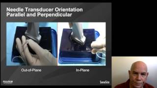
Ultrasound-guided Peripheral IV Access + 10 Common Pitfalls

Вставка
- Опубліковано 4 жов 2024
- A lecture describing the preparatory steps, 3 most common methods, and 10 common pitfalls of ultrasound-guided IV cannulation.
Bonus tip: During sniper technique, the depth of the target should match the needle-to-probe distance to triangulate cannulation.
www.us-guided.com. Laureano Andrade Vicenty, MD.


Very comprehensive tutorial! I greatly appreciate the time and effort you put into this presentation-from setup to pitfalls. Having practiced USIV cannulation over rhe past 5 years this video reminded me of some of my initial struggles. I'm sure this material will help others to gain mastery.
Best teacher ever.
Great video! I was having a hard time explaining some techniques to my trainees. This video has helped a lot!
Thank you! Shares are welcomed!
This is amazing!! Very useful video!
excellent
great video !!! Learned so much
Hello, great videos. Just out of curiosity, what IV brand/style do you use for ultrasound guided IVs? Where I work we currently use BD Nexiva 1.75" long IVs for US-guided IVs.
Thanks for sharing, very hard to find such practical tips. Quick question, why a 15 degree angle between skin and probe? Why not directly perpendicular ie 0 degrees?
Having a 15 degree probe-skin angle helps to reach that ideal 90 degree needle-probe angle for needle visualization.
Also thank you for your comment, please share with your colleagues 😊
@@TheMCATDisciplesI thought it might be so, but even in ur sniper technique for example the angle between ultrasound and needle is then 60 rather than 90 degrees. Does that pose a problem? Or to ask this question in another way what will the practitioner see if they maintain 0 degree probe angle/less than 90 degree ultrasound to needle angle?
Yes, the sniper technique is partially blind due to this 60 degree needle/probe angle, thus you will likely only see needle shadowing rather than a bright spot until you are within the vessel lumen, where the ultrasound wave conductivity improves due to the fluid (blood) within the vessel lumen.
@@TheMCATDisciples understood. thanks again for the video and for taking the time to reply
How is probe-to-skin angle 15 degrees? It’s clearly > 90 degrees according to picture… You mean probe-to-vertical plane angle 15 degrees, isn’t it?
Correct, 15 degrees from the vertical plane
Thank You for sharing but I have questions
What are your questions?
@@TheMCATDisciples *Hi* ( 1 ) Would you ever recommend placing a PICC Line without using any of these guides: a fluoroscope or ultrasound or CCVC? ( 2 ) What are the consequences of not using these guides?
( 3 ) For example, if one attempts to insert a PICC Line *into the left basilic vein* , but after placement of the sheath excessive bleeding occurs *because the PICC Line was actually placed into the artery in Error*
after realizing this it is decided let's hurry and remove this!............ (3 A) Is this a huge mistake? (3 B ) And what are some of the possible long term effects months or years later following this? *TY*
@@internationalintellectcrow2865 I can’t answer a question this specific over UA-cam.
If you have a question related to techniques in sonography, I would be happy to answer them.
@@TheMCATDisciples Sure is there a email to ask these questions?
?
fabulous video. But, why talk like a cowboy huffing smoke? Not like that in any other videos...