Currently in my Prelim-IM year, but will be starting Radiology in just a few months. Your videos are awesome! Really helped me out answering "pimp" questions during my Pulmonology rotation when the attending would pull up a CXR or Chest CT. Thank you for your time making these videos!
Thanks for video. Long Term (over 20 years) undiagnosed issues. Had a few of these scans. Incapacitated in the first 8 months, definitively started in left lung after a game of football. Notes of trapped air, and GGO, but it's plateaued. Will scrutinise my dicom set!
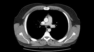
Very helpful to know a CT of chest is not just a 2 view pic, but a collection of images, like a tour of the inner organs full of info ready to be observed and reported on. Wow, yours is a step by step lesson, but only one suggestion: ask student at the beginning to note any n all observations, not wait til the end to say, "This is what I have been doing to write my impression," etc. Bravo.
Hey. Lovely video. Would be great if you could also add pointers as to how you anatomically delineate certain important structures. For eg. How does a beginner correctly identify the GE junction. Hope this could be done. Thanks again
Currently in a transitional year but starting rads next year. I have a lot of downtime on easy rotations so I’ve been watching your videos and then testing myself on E-anatomy. Hopefully I can start day 1 at least knowing the anatomy!
great video like that all other videos. great thanks to you dear Rishi. one day, can you make a video including your daily pracite. I mean in that video you dictating a few patient's thorax ct that containing some pathologies. mases, nodules, pneumonias, interstitial lung diseasess etc. I'm sure that you have a big portfolie of thorax pathologies. thanks, again.
I have this on my husband who has ipf I can’t tell anything compared to this. These are healthy lungs my husbands are moderate to severe ipf. I don’t know what I’m Looking for. Thanks explaining what I was looking at. It is what it is. Thanks. The pointer was hard to see
Lots of great information. I went to the ER and had a CT of my lungs looking for pulmonary embolism and it was clear. Is this type of CT the same as the one I had? I was concerned about my pancreas and was wondering if it was seen as well like in this scan.
I would also like to know the answer to this question please! Are all chesr CT scans like this? So if I got a chest CT scan to check for a PE kn the lungs specifically, would it also show the heart in great detail?
Hi, most chest CT scans don't go down far enough to see the entire pancreas. It will show the entire heart but not in great detail. Because the heart is beating while the scan is being done, the heart usually comes out blurry. There are special heart scans that can take a picture of the heart without blurring.
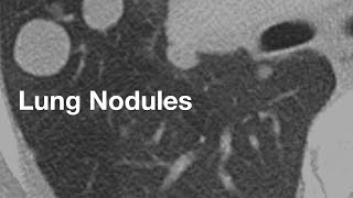
What can I do I have nodule in me lung is there anything I can do I have 8 goes of radiation it’s gone small but there other nodule in me lung also I got shadow on my lungs I need to have scan every 3 months
Great question. There is a wide variation in densities on CT from very dense (metals) to low density (air). But there are only so many shades of grey your eyes can perceive. When viewing a CT, there are lots of preset windows for certain tasks. For looking at the brain for stroke, for example, you want narrow windows that amplify the visual difference between grey and white matter. If you were to use wide windows that show calcium as white and air as black, the shades of grey will be spaced out too far so that subtle difference between grey and white matter will be lost.
Hello! Thanks for a useful video. What it can mean, when a doctor sees a lung nodule on the CT image, but this nodule is not visible on the lung window? Can it be some mistake?
if we found any lung lesion .....we will see this lesion on lung window or mediastinal window??????????????if first we see on lung window than what will tell us mediastinal window for that lesion????????
if it is small, you may not see it on mediastinal windows. if it is large enough, mediastinal windows can help you distinguish whether there is fat or calcium in the lesion.
Hi Kimon, thanks a lot. I'm not a cardiac imager, but check out this series by Methodist Hospital in Houston: ua-cam.com/users/HoustonMethodistDeBakeyCVEducationsearch?query=MRI
It is not a sufficient scan to detect breast cancer. Most regular CT scans of the chest are not sufficient for the heart. You need a CT specifically designed for the heart which synchronizes the scan to the heart beat. It is mainly for lungs and mediastinal structures, excluding the heart.o
A chest CT will show a good chunk of the liver. If the cirrhosis is advanced you can see it on a chest CT. But if it is in the early stages, you probably won't.
@@ThoracicRadiology Thanks for the reply.. i had right kidney cancer surgery years back and for the past at least 8 years i know of on my liver they keep calling this spot they see "Coarse or some saying dystrophic" calcifications in the upper right lobe of the hepatic dome depending on who reads them and even one scan saying -Again seen in the dome of the liver as the poorly enhancing foci corresponding to the dystrophic calcifications better seen on CT scan. i have had mri and ct scans am trying to find out what this means and what caused it if it's something to worry about ?scars from my cancer surgery? or because i have drink lots in the past, could it be cirrhosis? also should mention they said i have fatty liver and i also have cyst they seem to be inherited because my dad and a few of us have cyst if you could explain what could cause Coarse or dystrophic or poorly enhancing foci in the liver a little it be much appreciated :) worried it could be cirrhosis liver disease
Not sure the rates they are missed. But when they are small, like less than 1 cm, we can't always tell they are cancers. That's why we need a follow up study a lot of times. We rely on the growth to distinguish cancer from non cancer.
It can show abnormalities of the bone, yes. However some bone lesions and nearly all lesions of the spinal cord and the nerves coming off the cord are better evaluated by MRI.
First job in construction after working 12 years in fast food suppose to be carpentry my life was destroyed employer had me drilling in concrete and blowing out the holes with no protection and now I worry I will die with no hope no treatments no cure
If you are using the axial images, just scroll up to the first rib and count down until you find the rib that has the abnormality. Does that answer your question? Thx
I just had my CECT chest yesterday and my report says, "likely suggestive of complete radiological resolution of the disease". Can you please enlighten me what does this statement mean...?
I'm a medical dosimetry student. I'm presenting a lung cancer case this semester on a patient with severe bilateral emphysema. I wanted to give details on the appearance of emphysema on a CT image. Emphysema appears as a dark area in the lungs with absence of lung markings? Does it appear this way due to the thin lung tissue?
Hi Steve yeah it's because of a destruction of lung tissue and hyperinflation of the lungs leading to relatively greater air:tissue ratio compared to a normal person
Hi I got a ct heart scan today will this pick up anything else as am having serious issues with swallow my food and getting serious heart burn my esophagus is on fire
Hi, sorry, but without more information I can't really be sure what it is. It could be one of many different things, and the patient would need to talk to their doctor to figure out if it is significant and if so, what to do next.
Sir i did one hrct in 2020 other plain ct chest is 2023 now im woried about future cancer 2ct chest cause any cancer in my future pls help me im scared 🙏🏻😢
hi, the amount of radiation you received from those 2 scans is extremely low, and the potential for future cancer is either 0 or a number so close to 0 that we can't accurately measure it.
There are many types of chest scans and heart scans. If you are talking about CT, the heart scan will include part of the lungs too. Radiation is not usually an issue as most centers nowadays take precautions to reduce the amount of radiation to the amount just needed to achieve the diagnosis.
Hello hope everything is well. I have a question. Would a CT scan of the lungs show chemical damage or any microscopic damage to the lungs? I know it sounds dumb but im curious because i used to vape and so far the CT came clear, but im still worried
There is something called vaping-associated lung injury (VALI). This can be seen on CT, but it is usually associated with certain THC-based vape products that are manufactured with inferior ingredients. We saw a lot of cases in late 2019, but many of those products have been taken off the market, and we haven't seen many since. I glad you quit.
I am a beginner. If you could use pointers it would be great to understand. Honestly , I have no orientation of anatomy viewed through CT . I felt it was a bit fast as I could not place my concentration . However , great effort and good video .
hey :) thanks so much for the video! could you also explain how exactly you dictate? Like, do you only mention the pathological things or do you name every structure and say that its normal looking? i'm having real difficulties organizing my findings.. should i stick to my routine (top to bottom) or should i mention the important things first and then go on with the other ones? so confusing 😂
I use a template with the normal findings already in there and if there is something abnormal I replace that one thing with the finding that I see. The template that I use does not align exactly with my search pattern so I end up starting in the middle of my template, then the top, then bottom.
Thoracic Radiology sir I am not able to see video of right ventricular strain pulmonary embolism ..as u told in CT pulmonary embolism basics..that u will shortly update v
I have looked, but no I haven't found anything that is as good as osirix for free. I have heard people use 3d slicer before but I have not tried it myself.
Hi Rishi, I have a question here. I'm a CS student working in medical imaging. I'm about to do an application of deep neural networks for classification tasks in CT images. I have some CT data to work with, but I don't really know how to properly handle the data. I'm trying to sort slices related to the chest CT. Could you please reply to me with the timestamps of where exactly does the chest CT starts and ends (in the axial view)?
Hi, not sure if there is any good answer bc the timing is very dependent on the scanner. Some scanners can do a whole chest in like a couple seconds, others may take like 20 seconds or so.
Sir I had 2 HRCT chest for my COVID treatment..how much radiation do I got ( in MSV) ???? (CT values: 130 kv, 30mA, TI -4.3, GT-0.0, SL-0.6, W-350, C-50, 512 0/0,T20F SOPO 1, emotion 16 (2010) Sir is that low dose CT????
Dear Sir, I have been suferring from severe breathlessness on exertion after having corona. My SpO2 level at rest is fluctuating from 93 - 97%. It's comming down to 70% during walking. It has been six months since I contracted the deadly virus. I had my HRCT scan done but the doctors are telling it's normal. Please check my HRCT scan for any abnormalities and provide me with a path to get out of this suferring. Please help me Thanks
Sir mere bhai ne covid periyad me mahine 3bar HRCT karva liya tha abhi vo khud b tensan me he or puri family ko tensan me dala huva he use dar he muje cancer ho jayega 1st time riport normal tha fir b 2 bar HRCT karva ya totally 3 bar karvaya huva he sit pls riplay me use samja saku pls sir i need your advise
@@ThoracicRadiology Thanks! Reason is I have health anxiety pretty badly, and was concerned I have cirrhosis of liver. My doctor walked me through 1 of 2 chest CT's I had over the last few months for suspected PE and she reassured me I had no cirrhosis or damage of any kind (I also have had normal LFT for last 5 years) as she did not see any as a primary care doctor, and the radiologist did not note any liver abnormality in either scan. Radiologists are required to note such a incidental finding right?
@@nicholasdamuro1450 Yeah if a radiologist saw cirrhosis, they would report it. However, you can have cirrhosis of the liver and the liver can look normal on CT. CT is not the way to diagnose liver cirrhosis. If you had cirrhosis of the liver, you'd probably have LFTs that are abnormal and you would have yellow skin and yellow whites of your eyes.
Sir I had 2 HRCT chest for my COVID treatment..how much radiation do I got ( in MSV) ???? (CT values: 130 kv, 30mA, TI -4.3, GT-0.0, SL-0.6, W-350, C-50, 512 0/0,T20F SOPO 1, emotion 16 (2010) Sir is that low dose CT????
I can't tell from the information you gave me but it probably wasn't that much. To calculate the estimated effective dose take the DLP and multiply by 0.014.
@@ThoracicRadiology sir I'm not technical student..so i don't know how to find DLP. (they mentioned 10 cm length over there)...I can send you image of my film they given to me. Otherwise please tell me approximately how much dose they given in an average in HRCT chest in covid patients?? Because here in India Doctor advice average 2 CT scan.🙏
@@ThoracicRadiologyHere they only gave x-ray film and 1single page report...no any dosage information is mentioned...above values I found on a X-RAY film. that's only information I have.


Great video - I'm a stage IV renal cell carcinoma patient and this helped me understand my chest CT. Thank you for taking the time to make it.
Thanks Michael and good luck to you
How are you now?
@@masumiyet637 I don’t think he made it
Hallo how are you sir?
@@gauravjain7653 He has videos on his channel that were uploaded yesterday. He made it.
love your voice. It is really comfortable to listen.
Currently in my Prelim-IM year, but will be starting Radiology in just a few months. Your videos are awesome! Really helped me out answering "pimp" questions during my Pulmonology rotation when the attending would pull up a CXR or Chest CT. Thank you for your time making these videos!
Good luck with that rotation! If my residents watched these they would pretty much nail all my questions lol.
Wow! This was fascinating and it's amazing to think how technology has come so far. Thanks for posting these videos.
Thanks for video. Long Term (over 20 years) undiagnosed issues. Had a few of these scans. Incapacitated in the first 8 months, definitively started in left lung after a game of football. Notes of trapped air, and GGO, but it's plateaued. Will scrutinise my dicom set!
Very helpful to know a CT of chest is not just a 2 view pic, but a collection of images, like a tour of the inner organs full of info ready to be observed and reported on.
Wow, yours is a step by step lesson, but only one suggestion: ask student at the beginning to note any n all observations, not wait til the end to say, "This is what I have been doing to write my impression," etc.
Bravo.
thanks alot dear , iam prof in radiology from iraq . you did v much informative and respective .
Thank you. So helpful for a beginner
keep doing similar videos! 👌
Hey. Lovely video. Would be great if you could also add pointers as to how you anatomically delineate certain important structures. For eg. How does a beginner correctly identify the GE junction. Hope this could be done. Thanks again
Great suggestion! I'll think about this for a future video.
Currently in a transitional year but starting rads next year. I have a lot of downtime on easy rotations so I’ve been watching your videos and then testing myself on E-anatomy. Hopefully I can start day 1 at least knowing the anatomy!
Good luck!
You should have grasped your anatomy long while ago😂😂
Thank u very much sir..please keep uploading videos..can’t thank you enough…
Thank you so much 👍 it's of great help for a trainee learn the basics of ct chest ❤
You're welcome 😊
Great video, I reposted this video in my website with video summary that I made. Thank you!
Thank you so much!Very well explained and an excellent technique
So beautifully explained. !!
Excellent teaching presentation
great video like that all other videos. great thanks to you dear Rishi. one day, can you make a video including your daily pracite. I mean in that video you dictating a few patient's thorax ct that containing some pathologies. mases, nodules, pneumonias, interstitial lung diseasess etc. I'm sure that you have a big portfolie of thorax pathologies. thanks, again.
At 4:40 what is that round object to the right of the spine? ( Looks like a sphere.)
I think what you are looking at is the aorta
@@ThoracicRadiology Thank you.
Excellent video sir . Very well explained.
Sir any video about how to approach GGO on CT
Crystal clear, thank you a lot!
Actually amazing ❤
Very nice explanation
Can you please do a video on how to appreciate the heart chambers on non gated CT and how to tell if there is a dilatation or not ? TIA
Post rt changes noted in upper lobe of the lungs. It means
What do you mean by focal fibrotic changes in left lobe of the lungs
Excellent... signed a retired RN.
I have this on my husband who has ipf I can’t tell anything compared to this. These are healthy lungs my husbands are moderate to severe ipf. I don’t know what I’m
Looking for. Thanks explaining what I was looking at. It is what it is. Thanks. The pointer was hard to see
thanks and good luck to your husband.
How long does it take in real-time? Normal exam.
To read the study, a normal exam would probably take 10-15 minutes
Awesome video Sir.
thanks full showing inportnt work
Great work!
What was the radiation setting for a ct scan of chest with contrast?
Lymph node at 5:11 video how do u say its lymph node n not pulmonary thromboembolism ??
if you scroll up and down, you can determine that it is not in the vessel, but right next to the vessel.
Hi, thanks for this grest video. I just can't figure out one thing: linear opacity in the lung, like at 15:06, are blood vessels or airways? Regards
hi! the white linear opacities are blood vessels.
Watching so that i can understand cvs lectures better
Really simple and helpful
Lots of great information. I went to the ER and had a CT of my lungs looking for pulmonary embolism and it was clear. Is this type of CT the same as the one I had? I was concerned about my pancreas and was wondering if it was seen as well like in this scan.
I would also like to know the answer to this question please! Are all chesr CT scans like this? So if I got a chest CT scan to check for a PE kn the lungs specifically, would it also show the heart in great detail?
Hi, most chest CT scans don't go down far enough to see the entire pancreas. It will show the entire heart but not in great detail. Because the heart is beating while the scan is being done, the heart usually comes out blurry. There are special heart scans that can take a picture of the heart without blurring.
@@ThoracicRadiology But if they see anything suspicious they would note it?
Yes they would
Thank you very helpful and simple
thank you so much for this very informative and systematic video..i hope you can also make whole abdominal CT scan.
thanks for watching. I'm a chest radiologist so I'm probably not the best resource for abdominal CT stuff.
@@ThoracicRadiologydefinitely not hihi
What can I do I have nodule in me lung is there anything I can do I have 8 goes of radiation it’s gone small but there other nodule in me lung also I got shadow on my lungs I need to have scan every 3 months
شكرا جزيلا يا دكتور في ميزان حسناتك يارب
thank you so much, very useful
How to check by scan.. whether it is TB or not?
Thank you very much.
Awesome video
Would this test be enough to show a pulmonary embolism?
is it usual to use the word small instead of measurement because I am as a sarcoid patient they use this word to describe changes
Yeah if I use the word small instead of a measurement, it usually means that it is an insignificant finding.
thanks
Excellent video! I’m surgical resident. I have a question: What does “narrow the window” at 9:12 means? Thank you very much
Great question. There is a wide variation in densities on CT from very dense (metals) to low density (air). But there are only so many shades of grey your eyes can perceive. When viewing a CT, there are lots of preset windows for certain tasks. For looking at the brain for stroke, for example, you want narrow windows that amplify the visual difference between grey and white matter. If you were to use wide windows that show calcium as white and air as black, the shades of grey will be spaced out too far so that subtle difference between grey and white matter will be lost.
Oh, I got it! Thank you very much for replying!
Would this test show enlarged lymph nodes in the arm pit are? Armpit arc etc..
Yes it would
Hello! Thanks for a useful video. What it can mean, when a doctor sees a lung nodule on the CT image, but this nodule is not visible on the lung window? Can it be some mistake?
I'm not really sure that I understand the question.
if we found any lung lesion .....we will see this lesion on lung window or mediastinal window??????????????if first we see on lung window than what will tell us mediastinal window for that lesion????????
if it is small, you may not see it on mediastinal windows. if it is large enough, mediastinal windows can help you distinguish whether there is fat or calcium in the lesion.
High-yield presentation! Is there a chance for you to make some videos foucsing on cardiac MR? Looking forward to it.
Hi Kimon, thanks a lot. I'm not a cardiac imager, but check out this series by Methodist Hospital in Houston: ua-cam.com/users/HoustonMethodistDeBakeyCVEducationsearch?query=MRI
@@ThoracicRadiology Very helpful! Thanks a lot.
Sir is this scan significant for breast lung and heart collectively. Please guide about a detailed chest diagnosis thanks!
It is not a sufficient scan to detect breast cancer. Most regular CT scans of the chest are not sufficient for the heart. You need a CT specifically designed for the heart which synchronizes the scan to the heart beat. It is mainly for lungs and mediastinal structures, excluding the heart.o
@@ThoracicRadiology thanks sir you are doing great job👍.wish you more success .
Where do you practice right now?
@@ThoracicRadiology is there any specific name for the scan you recommended?
Excellent video! Great for a resident at the beginning! Hope you are gonna do more and more! greetings from Sicily :)
More to come!
how much of the liver can you see in a chest ct? can you diagnose or see cirrhosis in a chest ct?
A chest CT will show a good chunk of the liver. If the cirrhosis is advanced you can see it on a chest CT. But if it is in the early stages, you probably won't.
@@ThoracicRadiology Thanks for the reply.. i had right kidney cancer surgery years back and for the past at least 8 years i know of on my liver they keep calling this spot they see "Coarse or some saying dystrophic" calcifications in the upper right lobe of the hepatic dome depending on who reads them and even one scan saying -Again seen in the dome of the liver as the poorly enhancing foci corresponding to the dystrophic calcifications better seen on CT scan. i have had mri and ct scans am trying to find out what this means and what caused it if it's something to worry about ?scars from my cancer surgery? or because i have drink lots in the past, could it be cirrhosis? also should mention they said i have fatty liver and i also have cyst they seem to be inherited because my dad and a few of us have cyst if you could explain what could cause Coarse or dystrophic or poorly enhancing foci in the liver a little it be much appreciated :) worried it could be cirrhosis liver disease
Very helpfull
Very good video..shared on Radiology Vibes Facebook and telegram group.
How often are lung cancers missed
Not sure the rates they are missed. But when they are small, like less than 1 cm, we can't always tell they are cancers. That's why we need a follow up study a lot of times. We rely on the growth to distinguish cancer from non cancer.
@@ThoracicRadiologyI agree with buddy.. 😊
Thank you so much bro, from Bangladesh. Really overwhelming.
Thank you for watching!
So will chest ct scan show anything wrong with the spine and spine canal?
It can show abnormalities of the bone, yes. However some bone lesions and nearly all lesions of the spinal cord and the nerves coming off the cord are better evaluated by MRI.
Impressive thanks 👍
Thank you so much !
Brilliant!
First job in construction after working 12 years in fast food suppose to be carpentry my life was destroyed employer had me drilling in concrete and blowing out the holes with no protection and now I worry I will die with no hope no treatments no cure
Hello, how can we count ribs on ct scan? e.g if there is lyric lesion in right 5th rib then how we count the number ?
If you are using the axial images, just scroll up to the first rib and count down until you find the rib that has the abnormality. Does that answer your question? Thx
Nice sir
absolutely brilliant, thanks alot
I just had my CECT chest yesterday and my report says, "likely suggestive of complete radiological resolution of the disease". Can you please enlighten me what does this statement mean...?
It sounds to me like whatever you had went away.
I'm a medical dosimetry student. I'm presenting a lung cancer case this semester on a patient with severe bilateral emphysema. I wanted to give details on the appearance of emphysema on a CT image. Emphysema appears as a dark area in the lungs with absence of lung markings? Does it appear this way due to the thin lung tissue?
Hi Steve yeah it's because of a destruction of lung tissue and hyperinflation of the lungs leading to relatively greater air:tissue ratio compared to a normal person
Hi I got a ct heart scan today will this pick up anything else as am having serious issues with swallow my food and getting serious heart burn my esophagus is on fire
sometimes if a person has chronic heart burn (reflux), the esophagus can be thickened. that can be seen on a CT of the heart.
Thank you so so much! :)
What is meaning of nodular opacity noted in bilateral perihiller region.
Is it normal or not????
Hi, no that does not sound normal.
@@ThoracicRadiology please give me explain sir how it will effect our body and what should do for this. Chest X ray .
Hi, sorry, but without more information I can't really be sure what it is. It could be one of many different things, and the patient would need to talk to their doctor to figure out if it is significant and if so, what to do next.
Sir i did one hrct in 2020 other plain ct chest is 2023 now im woried about future cancer 2ct chest cause any cancer in my future pls help me im scared 🙏🏻😢
hi, the amount of radiation you received from those 2 scans is extremely low, and the potential for future cancer is either 0 or a number so close to 0 that we can't accurately measure it.
This was great Rishi! Just curious to know what WW and WL you use for interrogating the liver?
I use the manual window level tool to window it uniquely to each patient.
can a tumor still be detected with no contrast ct scan ?
Yes. Lung cancer screening is done without contrast
@@ThoracicRadiologyHi.. Should I get a HRCT or CECT Scan ? I want to rule out everything...
Hello, is a chest scan the same as a heart scan? Also is it too much radiation to do both?
There are many types of chest scans and heart scans. If you are talking about CT, the heart scan will include part of the lungs too. Radiation is not usually an issue as most centers nowadays take precautions to reduce the amount of radiation to the amount just needed to achieve the diagnosis.
Many thanks ☺
Hello hope everything is well. I have a question. Would a CT scan of the lungs show chemical damage or any microscopic damage to the lungs? I know it sounds dumb but im curious because i used to vape and so far the CT came clear, but im still worried
There is something called vaping-associated lung injury (VALI). This can be seen on CT, but it is usually associated with certain THC-based vape products that are manufactured with inferior ingredients. We saw a lot of cases in late 2019, but many of those products have been taken off the market, and we haven't seen many since. I glad you quit.
I am a beginner. If you could use pointers it would be great to understand. Honestly , I have no orientation of anatomy viewed through CT . I felt it was a bit fast as I could not place my concentration . However , great effort and good video .
Thx will keep in mind for future.
Great and concise.... The voice is too smooth like an nsdr video 😅
Thnku so much
Was this CT scan done with oral and IV contrast or none at all?
just IV contrast
Hello ! How did you do to have the coronal view thumbnail on the axial view when you are scrolling the series ? Thanks ! Great vieeo!
It comes up automatically with OsiriX. Do you have that program?
hey :) thanks so much for the video! could you also explain how exactly you dictate? Like, do you only mention the pathological things or do you name every structure and say that its normal looking? i'm having real difficulties organizing my findings.. should i stick to my routine (top to bottom) or should i mention the important things first and then go on with the other ones? so confusing 😂
I use a template with the normal findings already in there and if there is something abnormal I replace that one thing with the finding that I see. The template that I use does not align exactly with my search pattern so I end up starting in the middle of my template, then the top, then bottom.
Thoracic Radiology sir I am not able to see video of right ventricular strain pulmonary embolism ..as u told in CT pulmonary embolism basics..that u will shortly update v
This is great , thank you .
Quick question , do you know any program for windows like Osirix ?
I have looked, but no I haven't found anything that is as good as osirix for free. I have heard people use 3d slicer before but I have not tried it myself.
Hi Rishi, I have a question here. I'm a CS student working in medical imaging. I'm about to do an application of deep neural networks for classification tasks in CT images. I have some CT data to work with, but I don't really know how to properly handle the data. I'm trying to sort slices related to the chest CT. Could you please reply to me with the timestamps of where exactly does the chest CT starts and ends (in the axial view)?
Hi, not sure if there is any good answer bc the timing is very dependent on the scanner. Some scanners can do a whole chest in like a couple seconds, others may take like 20 seconds or so.
Sir I had 2 HRCT chest for my COVID treatment..how much radiation do I got ( in MSV) ????
(CT values: 130 kv, 30mA, TI -4.3, GT-0.0, SL-0.6, W-350, C-50, 512 0/0,T20F SOPO 1, emotion 16 (2010)
Sir is that low dose CT????
I have one question, I got a chest ct-scan for the first time , it is harmful for my body ? Please let me know, I am scared
No it is not harmful
Dear Sir,
I have been suferring from severe breathlessness on exertion after having corona. My SpO2 level at rest is fluctuating from 93 - 97%. It's comming down to 70% during walking. It has been six months since I contracted the deadly virus. I had my HRCT scan done but the doctors are telling it's normal.
Please check my HRCT scan for any abnormalities and provide me with a path to get out of this suferring.
Please help me
Thanks
Some people have difficulty breathing even with normal HRCT. CT can show some things but not everything. I would recommend seeing a lung specialist.
@@ThoracicRadiology Could you please check my HRCT Scan for any abnormalities if I send you the DICOM Image
Sir mere bhai ne covid periyad me mahine 3bar HRCT karva liya tha abhi vo khud b tensan me he or puri family ko tensan me dala huva he use dar he muje cancer ho jayega 1st time riport normal tha fir b 2 bar HRCT karva ya totally 3 bar karvaya huva he sit pls riplay me use samja saku pls sir i need your advise
If the CT is normal, no need to get more follow up CT. In my experience, COVID does not cause cancer. The risk of cancer from 3 CTs is negligible.
Thank you thank you thank you
Which software are you using sir ?
This is OsiriX (Mac only)
There is a small splenunculus
How long does this process take the average resident, and how long does it take you now that you’re experienced?
Me: usually 15 min. 10 min if completely normal. First year resident maybe 45 min. Fourth year, 20 min
Thanks
Is it typical to get as low as the kidneys and liver in a chest ct?
Yeah usually you get a good chunk of the liver and the very tops of the kidneys.
@@ThoracicRadiology Thanks! Reason is I have health anxiety pretty badly, and was concerned I have cirrhosis of liver. My doctor walked me through 1 of 2 chest CT's I had over the last few months for suspected PE and she reassured me I had no cirrhosis or damage of any kind (I also have had normal LFT for last 5 years) as she did not see any as a primary care doctor, and the radiologist did not note any liver abnormality in either scan. Radiologists are required to note such a incidental finding right?
@@nicholasdamuro1450 Yeah if a radiologist saw cirrhosis, they would report it. However, you can have cirrhosis of the liver and the liver can look normal on CT. CT is not the way to diagnose liver cirrhosis. If you had cirrhosis of the liver, you'd probably have LFTs that are abnormal and you would have yellow skin and yellow whites of your eyes.
Do you know, why there is always a big right hilar lymphnode ?
Not sure, but I think that it is prob something to do with the way the hilum is oriented.
Nice
Sir I had 2 HRCT chest for my COVID treatment..how much radiation do I got ( in MSV) ????
(CT values: 130 kv, 30mA, TI -4.3, GT-0.0, SL-0.6, W-350, C-50, 512 0/0,T20F SOPO 1, emotion 16 (2010)
Sir is that low dose CT????
I can't tell from the information you gave me but it probably wasn't that much. To calculate the estimated effective dose take the DLP and multiply by 0.014.
@@ThoracicRadiology sir I'm not technical student..so i don't know how to find DLP. (they mentioned 10 cm length over there)...I can send you image of my film they given to me.
Otherwise please tell me approximately how much dose they given in an average in HRCT chest in covid patients?? Because here in India Doctor advice average 2 CT scan.🙏
Our average here is like 3-5 mSv for 1 low dose CT. You can find the DLP in the radiation dose page of your scan.
@@ThoracicRadiologyHere they only gave x-ray film and 1single page report...no any dosage information is mentioned...above values I found on a X-RAY film. that's only information I have.
@@theCalmMonk how r u now ? Ur age?
Amazing..thank you
Thank you Atefeh!
@@ThoracicRadiology I am aspiring to be a CT surgeon and sadly I use to mistake a lymph node or a PE
softwar name please???
Osirix
@@ThoracicRadiology can you send me software link please???