In order to make high quality content consistently, we need support from you. Please support us by using super thanks option. Super thanks icon is present below the video ( a heart sign with $ in it ) . You can support using paytm/ phone pe/ gPay / paypal. Your small contribution means a lot for us.
Could you please help me by sharing my contents with your friends group/ college group. I put huge efforts in making these videos but unfortunately not a lot of people are watching this.
Very niche topic of cell biology. Glad you made a video explaining it here! A question however - The 4 quadrants we see in the result of FACS experiments. How are they named I, II, III and IV. Which one is labelled what? I ask this as there's a question in CSIR 2012 asking about the quadrant that early apoptotic cells are found in? The answer is Quadrant III but i am curious what the other quadrant names are, like the one on the upper left, is it Quad I or something else? Thanks! 👋😄
In order to make high quality content consistently, we need support from you. Please support us by using super thanks option. Super thanks icon is present below the video ( a heart sign with $ in it ) . You can support using paytm/ phone pe/ gPay / paypal. Your small contribution means a lot for us.
@@animatedbiologywitharpan Thank u so much Sir... Plz make them if possible... Your explanation is super and totally different from other videos... God bless you...
Could you please help me by sharing my contents with your friends group/ college group. I put huge efforts in making these videos but unfortunately not a lot of people are watching this.
Generally, cells are more stable and tolerate stress better when they're cold. So before FACs ( which is quite stressful for cells) cells are kept on ice.
Hi, I usually trypsinize the cells and collect them before treating the cells with the annexin V-PI stains, then it that sense do you think I should also collect the floating cells in the old media together with the trypsinized cells? Then may i also know how you gate them 4 quadrants in the end? Using a positive control that give you different cell death regions with single stain? Like PI-only or Annx V-only
Really glad to know it was useful. Please share my channel with friends. You can support the channel by clicking on the super like icon below the video ( a heart sign with $ in it ) . You can support using paytm/ phone pe/ gPay / paypal. Your small contribution means a lot for me
These overall videos gives an concept and some understanding about the process. It's not a protocol video which will describe each and every nitty-gritty details
Thanks alot very good explanation. I have question The last part of the diagram how can we know that, for example this colours specefied for each part? What is the meaning of the red colour, yellow, blue...
Annexin A5 is involved in inhibition of blood coagulation by competing for phosphatidylserine binding sites with prothrombin and also to inhibit the activity of phospholipase A1. So from a structural point of view it is more likely to bind PS and not PC or PE.


Very clear explanation. No stammering and just zipped through and crisp with good fig. Thank you.
In order to make high quality content consistently, we need support from you. Please support us by using super thanks option. Super thanks icon is present below the video ( a heart sign with $ in it ) . You can support using paytm/ phone pe/ gPay / paypal. Your small contribution means a lot for us.
I actually love you.. by far the best explanation for this I have seen! I suddenly feel less stupid, Thanks :)
Could you please help me by sharing my contents with your friends group/ college group. I put huge efforts in making these videos but unfortunately not a lot of people are watching this.
Your videos helped me to clear DBT BET. Thank you so much.
In order to make high quality content consistently, we need support from you. Please support us by using super thanks option. Super thanks icon is present below the video ( a heart sign with $ in it ) . You can support using paytm/ phone pe/ gPay / paypal. Your small contribution means a lot for us.
Great video! Is there a need for a wash step after the Annexin/PI incubation?
precise content..thank you
Please share my channel link with your friends and help me to reach big audiance
Best explanation ❤
Could you please help me by sharing my contents with your friends group/ college group. I put huge efforts in making these videos but unfortunately not a lot of people are watching this.
Excellent video for understanding.
Please share my channel link with your friends .
Your videos are so helpful for me, thank you!
Please share my channel link with your friends and help me to reach big audience
Thanks for making this video, it is really helpful. Can you please make videos on other cell death assays like ferroptosis and pyroptosis?.
Thanks for your suggestion….I will try
This was so easy to understand. Thank you for posting😊
Please share my channel link with your friends and help me to reach big audiance
Very niche topic of cell biology. Glad you made a video explaining it here!
A question however - The 4 quadrants we see in the result of FACS experiments. How are they named I, II, III and IV. Which one is labelled what? I ask this as there's a question in CSIR 2012 asking about the quadrant that early apoptotic cells are found in?
The answer is Quadrant III but i am curious what the other quadrant names are, like the one on the upper left, is it Quad I or something else? Thanks! 👋😄
I will answer this in my other channel by making a explainer video. I will post the link here
Very good explanation
In order to make high quality content consistently, we need support from you. Please support us by using super thanks option. Super thanks icon is present below the video ( a heart sign with $ in it ) . You can support using paytm/ phone pe/ gPay / paypal. Your small contribution means a lot for us.
Thanks a lot ! very helpful !
Please share my channel link with your friends and help me to reach big audiance
Thank you so much for this video.✨
Please share my channel link with your friends and help me to reach big audience
HI this is very simplified explanation, i love it, i have a questuon is it to possible use of Annexin-V PI to access pyroptotic cells?
Most likely no
Thank you so much Sir.. Nice explanation... Sir could you make new videos on AO/EB and DAPI staining
These are stains that binds to dna , I will try to make a video if I get some time
@@animatedbiologywitharpan Thank u so much Sir... Plz make them if possible... Your explanation is super and totally different from other videos... God bless you...
Best video
Could you please help me by sharing my contents with your friends group/ college group. I put huge efforts in making these videos but unfortunately not a lot of people are watching this.
may i ask for the reason why will need to keep the cells with staining solution in ice?
Generally, cells are more stable and tolerate stress better when they're cold. So before FACs ( which is quite stressful for cells) cells are kept on ice.
@@animatedbiologywitharpan thank you so much
@@白馬義從寶葵 please support this channel (even 1$) using super thanks. Super thanks in a heart shape icon below the video.
Hi, I usually trypsinize the cells and collect them before treating the cells with the annexin V-PI stains, then it that sense do you think I should also collect the floating cells in the old media together with the trypsinized cells? Then may i also know how you gate them 4 quadrants in the end? Using a positive control that give you different cell death regions with single stain? Like PI-only or Annx V-only
Very good video thank you. But why can't Annexin V connect with the membran in Necrosis?
Because phosphatidyl serine is not flipped on the outer leaflet. And Annexin selectively binds to phosphatidyl serine
@@animatedbiologywitharpan This makes sense. Thank you 😊
What should be the final conc of PI for FACS?
Need ur help in solving immunology questions
Perfect
Really glad to know it was useful. Please share my channel with friends. You can support the channel by clicking on the super like icon below the video ( a heart sign with $ in it ) . You can support using paytm/ phone pe/ gPay / paypal. Your small contribution means a lot for me
Discard media and then wash with PBS and then add annexin binding buffer? I mean no need to scrape the cell or trypsinise ?
These overall videos gives an concept and some understanding about the process. It's not a protocol video which will describe each and every nitty-gritty details
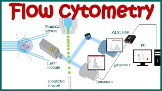
Thanks alot very good explanation. I have question
The last part of the diagram how can we know that, for example this colours specefied for each part? What is the meaning of the red colour, yellow, blue...
Thats a density heatmap. Color code: Hotter color ( redish) = high cell density, cooler color ( bluish) = low cell dnsity
@@animatedbiologywitharpan many thanks
@@za7607 please share my channel link with friends
Why annexin not binds to ps and pc they are outside of the membrane only know .and why annexin specifically binding with ps rather than ps and pc
Annexin A5 is involved in inhibition of blood coagulation by competing for phosphatidylserine binding sites with prothrombin and also to inhibit the activity of phospholipase A1. So from a structural point of view it is more likely to bind PS and not PC or PE.