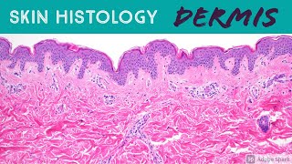
Skin Histology: Epidermis Layers (stratum basale, spinosum, granulosum, lucidum & corneum)

Вставка
- Опубліковано 7 лют 2025
- Excerpt from my Normal Skin Histology video: kikoxp.com/pos....
A complete organized library of all my videos, digital slides, pics, & sample pathology reports is available here: kikoxp.com/pos... (dermpath) & kikoxp.com/pos... (bone/soft tissue sarcoma pathology).
Please check out my Soft Tissue Pathology & Dermatopathology survival guide textbooks: bit.ly/2Te2haB
Also, in the past I used "keratinocyte" and "squamous cell" interchangeably (this is because in dermatopathology, we see and talk about squamous cell carcinomas all the time, and those tumors are composed of keratinocytes). But technically, in normal skin histology, "squamous cell" refers only to the flattened keratinocytes in the superficial epidermis. Thankfully, a histology PhD colleague pointed this out to me and corrected my lazy nomenclature!
This video is geared towards medical students, pathology or dermatology residents, or practicing pathologists or dermatologists. Of course, this video is for educational purposes only and is not formal medical advice or consultation.
Presented by Jerad M. Gardner, MD. Please subscribe to my channel to be notified of new pathology teaching videos.
Follow me on:
Snapchat: JMGardnerMD
Twitter: @JMGardnerMD
Instagram: @JMGardnerMD
Kiko: kikoxp.com/pro...
Facebook: / jmgardnermd


Excerpt from my Normal Skin Histology video (full video here): kikoxp.com/posts/3660. A complete organized library of all my videos, digital slides, pics, & sample pathology reports is available here: kikoxp.com/posts/5084 (dermpath) & kikoxp.com/posts/5083 (bone/soft tissue sarcoma pathology).
If it would be possible could you please show in a video how to orient about the normal difference in size among the cells in a tissue and also the different types of magnifiers of the microscope and the dimension in the field. For example HPF at 40 x represents 1 mm?
It becomes paramount to know the normal histology very well first in order to identify what is abnormal. Great correlation with diseases and IHC!!!! Fabulous
your enthusiasm made this awesome, instructive video extra great! thank you for walking thru real slides, so so helpful!
Incredibly helpful video
Loved every bit of the video ❤❤thank you
Awesome video! Just what I needed for my test.
Excellent .Thanks a lot dear for sharing this video.
Excellent video. Thank you very much
Dose it have treatment or cream and soap to clear it
Thank you that saved me
Thank you very much