Structure Of The Skin - Layers Of Skin - Types Of Skin - Types Of Skin Cells - Integumentary System

Вставка
- Опубліковано 16 чер 2024
- In this video we discuss the structure of the skin, we look at the 2 different layers of skin, the epidermis and the dermis, the structure of each of these layers, and the 2 different types of skin, thick skin and thin skin.
Transcript/notes
Structure of skin
Human skin, which is also called the integument or the cutaneous membrane, is made up of 2 layers, the epidermis and the dermis, which are labeled on this model of skin. The subcutaneous layer at the bottom has also been labeled, however, it is not actually a part of the structure of skin, but it is connected to the dermis of the skin.
Let’s start by looking at the epidermis. The epidermis consists of 4 to 5 layers depending on the type of skin. Thick skin has 5 layers, and it is found in the palms of the hands, and on the soles of the feet. Thin skin has 4 layers and is what covers most of the body. The skin model we are looking at has all 5 layers.
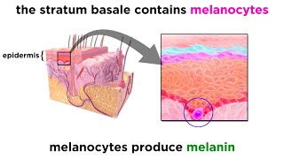
The bottom or deep layer is called the stratum basale. It is made up of a single layer of cells attached to a basement membrane. There are 3 types of cells in the stratum basale; keratinocytes, melanocytes and tactile cells.
Keratinocytes are the most common cell in this layer, and they go through cell division to replace cells that are shed from the surface of the skin. These cells can produce a tough structural protein called keratin which strengthens the skin and makes it almost waterproof.
Melanocytes are scattered among the keratinocytes and they produce the pigment melanin in reaction to exposure to ultraviolet light. Melanin gets transferred to keratinocytes and surrounds the nucleus to protect DNA from mutating from ultraviolet radiation.
Tactile cells are also scattered among the keratinocytes, and they serve as light touch receptors.
The next layer, moving upwards in the epidermis is the stratum spinosum. This layer is made up of daughter keratinocytes made from dividing cells in the stratum basale layer below, and epidermal dendritic cells. The daughter keratinocytes connect to neighboring cells desmosomes, which are one of the ways cells connect to one another, giving them a prickly appearance. The dendritic cells are immune cells that help fight infections in the skin.
Moving upwards, the next layer is the stratum granulosum. This layer is comprised of 3 to 5 layers of keratinocytes. The process of keratinaztion begins in this layer of the epidermis. Keratinization is where the keratinocytes fill with the keratin protein metioned earlier. This process continues as the cells move upwards in the epidermis, and as it continues, the cell’s nucleus and organelles are eliminated and the cell dies.
The next layer up is the stratum lucidum. This layer is only found in the thick skin in the palms and soles of the feet. The keratinocytes in this layer are clear, flat, closely packed and have no nucleus or organelles. They are also filled with a protein called eleidin, which is eventually transformed into keratin.
The last or top layer is called the stratum corneum. This layer is comprised of dead keratinized cells. It takes about 2 weeks for a new keratinocyte to reach the stratum corneum, and it remains in this layer for about another 2 weeks before it is shed.
Now let’s look at the dermis. The dermis is comprised of connective tissue proper with collagen being the most plentiful type of fiber found throughout the dermis. The dermis also houses other structures such as blood vessels, hair follicles, sweat glands, sebaceous glands, which secrete a lubricating oil, sensory nerve endings, nail roots and arrector pili muscles which affect hair follicles.
The dermis has 2 layers; a papillary layer, and a reticular layer.
The papillary layer is the top superficial layer of the dermis, it is composed of loose connective tissue and forms bumps or projections called dermal papillae that fit with the epidermal ridges of the epidermis. The form of the dermal papillae and epidermal ridges increases the surface area of contact between the 2 layers. The dermal papillae contain capillaries that supply nutrients to the cells of the epidermis, and they contain sensory nerve endings that help monitor touch on the surface of the skin.
The reticular layer is composed of dense connective tissue and it extends from the papillary layer to the deeper subcutaneous layer. It is composed of a dense connective tissue with some elastic fibers and many bundles of collagen fibers.
Timestamps
0:00 Overview of the structure of skin
0:28 The epidermis
0:49 Stratum basale
1:03 Keratinocyte cells
1:18 Melanocyte cells
1:34 Tactile cells
1:44 Stratum spinosum
2:09 Stratum granulosum
2:36 Stratum lucidum
2:56 Stratum corneum
3:11 The dermis
3:46 The papillary layer
4:17 The reticular layer
4:28 The subcutaneous layer


Very good overview. One of the best that I have seen. Concise and clear explanations. Thanks.
Aghh thank god ive found the most benificial vid ever really helps alot in my exam and i got 57/60 because of this vid.keep uploading more educational vids especially science
Thank you sir, keep making videos like this, this is not only helpful for exam but also for general information.
Great, great, great explanation.
Keep making videos like this. This made me understand more for my exam.
Great to hear the video helped you out Mia, thanks for watching.
U r awesome
Please do like these videos. U r a good lecturer
Very helpful!
Thanks it’s really helpful 🙂.
Really good explanation.
THANK YOU!!!
Best video 👌♥️
Great refresher for my final exam
The most helpful and beneficial video thank you sooooo much
You are welcome Shamama Behzad.
thank you so much , your explanation make me understand very well this chapter 5
You are welcome Vipon Kamango, and thanks for watching.
Thank u for this. Itso so easy to learn by watching videosthan reading
Awesome
man I didn’t watch this for no exam I watched it because I love my cells.
I need this for my exams
Fantastic illustration sir. The content is exactly according to my syllabus. Please make more and more videos. I love your teaching method. The only thing I want in your videos is the slow pace. So, its a request to be somewhat slow while explaining. Overall, awesome job. Keep it up.
Thanks!
Thanks for watching Anmol Dhillon, and thanks for the feedback.
Thanks you so much 😇 this is hope me a lot as I now doing enrolled nursing in Australia 🇦🇺
Thanks for sharing that Lydia and good luck with your studies.
way much better than books
It's helpful to me for my exam 😀😀😀
Thank you very much!...wow!🙌👏👏
You are welcome, thanks for watching.
@@whatsupdude2778 ну со ту камес кар кирло ту бэш по кар
I am skin. Thank YOUUUU!!!😆☕🥁🌉
Is it keratinocytes or epithelial cells which divide n undergo keratinization? In skin... i heard its epithelial cells which undergo keratinization n form lipids.. then move upwards? Im confused....
Thank you soooo much.... :))))*it is really helpful for my examination
Also helpful for icse examination
Good video
Thanks my friend.
Wow I love this
Thanks dude, where are u from brother,I am saying becos if I need any help or I get any doubt can I cmnt or not
I am frist person watch
so what is the function of each layer?
thank you x 1000000000000000000000000000000000000000000000000
Thx boi student
thank you for this! the book only showed superficial info and ddin't tackle specific cells :'((( thank youuu!!!
You're so welcome!
Hey my dood can you do a video about the reproduction system cause ur good at explaining things :)
My friend got in accident so I watch this because u can see the bone clearly
La neta quien vino aquí por la ms Daniela😂🤷🏼♂️
I am a huge fan of your videos but for some strange reason i had a hard time with this video because of the fact it was not animated.
Hey Nafisa101, I think you are correct. I thought it might be easier for people to understand using a skin model, but looking back at the video, I probably should have just stuck to animating it. Thanks for the feedback, and thanks for watching.
Whats Up Dude your welcome
I watched so I can figure out where to body stitch, I have no idea how to do it safely
.