Hi! Join to help me create more free educational content 🫀Join: ua-cam.com/channels/Er7pkSXVsHcBLLBcJAGV-Q.htmljoin 📸Instagram: instagram.com/taimtalksmed/
Never Mind Mistakes WOW WOW What A Lot Of Hard Work And Time You Put Into These Beautiful Animated Videos You A A Genius A Super Star And A Super Wonderful Teacher Thank You So Much For Sharing Your Beautiful Wonderful Knowledge And Time And Patience And Teaching and Lots Of Hard Work Put Into These Beautiful Videos @ Taim Talks Med 👍👍❤️❤️
This is amazing, honestly every other anatomy source was melting my brain and I understood everything here, explained simply yet detailed. Defo subbed!
@@TaimTalksMed if it is possible, could u tell me that where did u study and where r u from yrself. Which books can u recommend us to study anatomy? I would be glad if u reply me! Sincerely, Diyorbek from Tajikistan.
I am so grateful i found your channel. You made me understand neuroanatomy completely. Thabk you do much for all you do. You deserve more followers ❤❤❤
Thank you very much for the clarification, I find it extremely useful, such explanations are never enough. Thank you very much and I hope to deliver many others
omg I m just so happy that I was able to find these videos but sad too as I already wasted my time with Kaplan ( made me so sleepy )…this was so quick n detailed and discussing the tracts at the same time ,keeping neurology interesting and didn't made me tired or procrastinate, cant thank u enough …😄😃 well almost forgot to mention the details u have written with your videos n also mentioning the time n topic saved a lot of time if i wanna jump to certain section of the video
First off, thanks so much for your videos, they are all really informative and so well made! However I have one question here: At about 13:40, you talk about the olivocerebellar tract and how it comes form the olivary nuclei and then goes on into the cerebellum. Wouldn't this be a sensory/ascending pathway and thus blue? Hoping you will see this and answer me because it confuses me a little! Thank so much!😃
Totally understand your confusion! Keep in mind that the olivocerebellar pathway initially relays information from the cerebral cortex that projects to the olivary nuclei and then to the cerebellum. So if you make a ''mindmap'' of this pathway, you'll see that it is in fact descending. You can blame me for not elaborating on this one =)
Wow great video you inspired me this is Jerusalem the cave of Brahma the holy of holies the Ark of the Covenant Golgotha and it's in each of us how wonderful
hey at the end at min 16:19 u switched the names of cuneate nucleus and gracile nucleus ? and u mentioned in the video when u were explaining them other way around
Thank you for a very good explanation of the structure and function of the medulla. I have several autoimmune diseases and had inflammation in this portion of the brain as a consequence. It is helpful in explaining some of my symptoms, wish there were better methods for helping these symptoms!
Sorry to hear about your condition, and I truly hope they find better treatment options in the future as well! I'm glad I could contribute to your learning =)
Hey man! Glad you're finding the videos helpful! Pons is up on Sunday, 4th ventricle and rhomboid fossa on the 15.09. I'm sure you'll do great in the test, just keep your head cold and focused 💪
are pyramidal & medial lemininsci occur at vertical or transverse plane? in another way : do the two decussations actually appear in a transverse section ?
Deccusations in general can be seen in both planes. You'd rather visualize these tracks using visual imaging like track density imaging and tractographies though (as it's hard to localize tracts in the white matter on a cadaver). I only show relative schemes though, really only to help with the visual learning. Hope you found the video helpful nevertheless 😊
I did remake all the schemes at different levels of the brainstem, but they will be available on PDF notes as soon as I am done with all the topics in Anatomy
Glad it helped! Yeah the UA-cam algorithm's not my friend unfo😅 The more people reacts with the video, the higher it gets in the search results though; so I'm staying positive! 💪
@aditeecreationsisreal it's just the number on the table at the end that is switched, I made a comment about it. The explanation part is correct 😊 I haven't corrected the video, too minor mistake to reupload
guys i dont understand the kind of fibres that goes to the gracilis nucleus,i understand that there coming from the lower part of the body but what is propreception what kind of fibres are they ???
Blessings in an abundance of the faith. I salute you. 😘🙏Grace and peace be unto you and to this place. God bless you. 🤍🌈🤍Thank you. 💌Will you believe and receive Jesus Christ as your LORD and personal saviour??? I do. 😊


Hi! Join to help me create more free educational content
🫀Join: ua-cam.com/channels/Er7pkSXVsHcBLLBcJAGV-Q.htmljoin
📸Instagram: instagram.com/taimtalksmed/
PS! There's a little mistake in the table at the end (grey matter). Number 2 and 3 are switched. Apologize for the minor mistake!
There is a mistake only in 2 and 3 right ??
Rest of the table is correct right??
Not a problem 😘😘 it really helped a lot
Never Mind Mistakes WOW WOW What A Lot Of Hard Work And Time You Put Into These Beautiful Animated Videos You A A Genius A Super Star And A Super Wonderful Teacher Thank You So Much For Sharing Your Beautiful Wonderful Knowledge And Time And Patience And Teaching and Lots Of Hard Work Put Into These Beautiful Videos @ Taim Talks Med 👍👍❤️❤️
Very helpful thanks...what gaoodns when someone has a mass in the medulla?
This is amazing, honestly every other anatomy source was melting my brain and I understood everything here, explained simply yet detailed. Defo subbed!
For every lesson of anatomy, I watch your videos. They are amazing and easily instructed.
(From Uzbekistan 🇺🇿)
I'm stopping midvideo just to say thank you so much, you make it so much easier to understand and memorise!
Ur videos are by far the best neuroanatomy videos I've come across on UA-cam!
This is so detailed, thank you! Hope your channel grows much more bro!
Thank you so much!!
The best video I found to get a good idea of the medulla as well as a brief understanding of the conductive system
All your videos are too available for every medstudents. May God blesses you. Thanks so much for your aid
Knowledge should be free, In my opinion! Glad I could help you through these videos 😊
@@TaimTalksMed if it is possible, could u tell me that where did u study and where r u from yrself. Which books can u recommend us to study anatomy?
I would be glad if u reply me!
Sincerely, Diyorbek from Tajikistan.
How am I just seeing this channel... The best detailed yet easy to understand explanation... Kudos 👏
Happy to hear! 😊
This video is very detailed , well explained and simple. It has really helped me in my studies. Thank you and keep up the good work .
Glad to hear! All the best with your studies :)
شكراً دكتور🤍🤍.
الله يزيد علمك ويبارك فيه🤲🏻🤍.
amazing content.honestly without you I can never graduate from med school
I am so grateful i found your channel. You made me understand neuroanatomy completely. Thabk you do much for all you do. You deserve more followers ❤❤❤
You are a better teacher than all doctors in iraq , thank you very much
I wish I can like this video a million times
this video has done so much for me in such little time. Thank you🙏🏾
Thank you very much for the clarification, I find it extremely useful, such explanations are never enough. Thank you very much and I hope to deliver many others
Thank you! Appreciate the kind words
Best explanation I have ever seen!!
thank you this was exceptional !!!
thank you teacher, it's very helpful and easy to understand, from viet nam
Very full and easy-to-follow video. Awesome work
I finally understand. Thank you 😭
The best explanation , it’s realllly benefit me ❤❤❤ thanks man ❤❤❤
omg I m just so happy that I was able to find these videos but sad too as I already wasted my time with Kaplan ( made me so sleepy )…this was so quick n detailed and discussing the tracts at the same time ,keeping neurology interesting and didn't made me tired or procrastinate, cant thank u enough …😄😃 well almost forgot to mention the details u have written with your videos n also mentioning the time n topic saved a lot of time if i wanna jump to certain section of the video
Nice!💪🏽Glad to hear! 🙂 All the best with your studies
Nice video i had ever seen on medulla just in 16 minutes..thnku so much sir..😊
WONDERFULL! in only 16 mins…
Great videos, I love how you breakdown connections between structures. Please keep making more
Thank you. You are doing best🎉🎉
It's the best vedio about brain stem anatomy
First off, thanks so much for your videos, they are all really informative and so well made! However I have one question here: At about 13:40, you talk about the olivocerebellar tract and how it comes form the olivary nuclei and then goes on into the cerebellum. Wouldn't this be a sensory/ascending pathway and thus blue? Hoping you will see this and answer me because it confuses me a little! Thank so much!😃
Totally understand your confusion!
Keep in mind that the olivocerebellar pathway initially relays information from the cerebral cortex that projects to the olivary nuclei and then to the cerebellum. So if you make a ''mindmap'' of this pathway, you'll see that it is in fact descending.
You can blame me for not elaborating on this one =)
@@TaimTalksMed Ah I see that makes sense!! Thanks so much for your very quick answer and explanation! Got it 😃
You're a legend. Thanks for being the only reason I pass neuroanatomy.
Thank you very much 😭you help me to understand things 2 month I try to understand
Thank you sir 🙏it's very helpful for us to understand in 3d way
Amazing amazing amazing 💛💛💛💛
3 hours dr najeeb
After I get this good visual revision
Man you did BEST GOOD DEED you could do thanks
You're welcome! Happy to hear it's been helpful
بارك الله لك في علمك اخي ❤️
Exceptional video!
This really helped me a lottt
Thankssss 🌹
Amazing 🤩 thank you very much
Wow great video you inspired me this is Jerusalem the cave of Brahma the holy of holies the Ark of the Covenant Golgotha and it's in each of us how wonderful
Very very good videos💙
that was so greattt and easy to understand thanks alottt😭🔥🔥🔥🔥🔥🔥
You're welcome! Glad it helped
We wish you the highest levels
Thank you ❤🎉 this video faciliate everything
TQ very much for valuable information
hey at the end at min 16:19 u switched the names of cuneate nucleus and gracile nucleus ? and u mentioned in the video when u were explaining them other way around
Thank you for pointing that out! I've pinned a little note in the comment section :)
Best video❤❤❤❤❤
تحفة تحفة تحفة شرحك بكددد جامممد جدااا
متشكرة حضرتك 😭❤️
Actually Your explanation is so great, you saved me.
Thank you soooooooooooooo match ❤️
Really glad to hear! 💪🏽🥳💝
All the best with your studies!
This is amazing,your explanation made this very easy.Thank you:)
Excellent explanation, it helped me a lot Thank u so much 👏🏼
Awesome vid thanks a lot
Thank you so much, this is very veryyy helpful 😊
Interesting ,detailed eaiser to grab 🤗
Great videos, thank you so much 👍👍👍
Fabulous sir......
Спасибо ❤
Amazing
keep making more.
Thank you for a very good explanation of the structure and function of the medulla. I have several autoimmune diseases and had inflammation in this portion of the brain as a consequence. It is helpful in explaining some of my symptoms, wish there were better methods for helping these symptoms!
Sorry to hear about your condition, and I truly hope they find better treatment options in the future as well! I'm glad I could contribute to your learning =)
thanks😆😁😁😃😀
So nice explaine
Thank you❤❤❤❤
You're welcome! Glad you found it helpful =)
God bless you!
Very precise!
Awesome ...keep your hard working for sure you will reach !🙂❤️
But dont sell the date cause I cant buy them 😅😂
Please upload pons,midbrain and reticular formation have my last attempt on the 17th September 😭
Hey man! Glad you're finding the videos helpful! Pons is up on Sunday, 4th ventricle and rhomboid fossa on the 15.09. I'm sure you'll do great in the test, just keep your head cold and focused 💪
Nice presentation
In which video do we have the internal structure of inferior cross sectioned part of medulla oblongata ?
are pyramidal & medial lemininsci occur at vertical or transverse plane?
in another way : do the two decussations actually appear in a transverse section ?
Deccusations in general can be seen in both planes. You'd rather visualize these tracks using visual imaging like track density imaging and tractographies though (as it's hard to localize tracts in the white matter on a cadaver). I only show relative schemes though, really only to help with the visual learning. Hope you found the video helpful nevertheless 😊
Amazing!
This is really impressive and helpful video. Thank you for this
Thank you!
Veryy coool video!
Beautiful explanation!
وما تشائون الا ان يشاء الله رب العالمين
Thanks much bro
Love this
good dedication . keep it up. u r bit fast. try to tune for best speed..
Is there a vd of urs at diff lv of dissections of medulla oblongata ?
I did remake all the schemes at different levels of the brainstem, but they will be available on PDF notes as soon as I am done with all the topics in Anatomy
@@TaimTalksMed okie thank u
Thank you
wonderful explanation. only if it shows first when searching :l
Glad it helped! Yeah the UA-cam algorithm's not my friend unfo😅 The more people reacts with the video, the higher it gets in the search results though; so I'm staying positive! 💪
Is the mistake already corrected in the video?? Because gracile is for lower limb , and you wrote the same but the comments says its wrong
@aditeecreationsisreal it's just the number on the table at the end that is switched, I made a comment about it. The explanation part is correct 😊 I haven't corrected the video, too minor mistake to reupload
best 👍
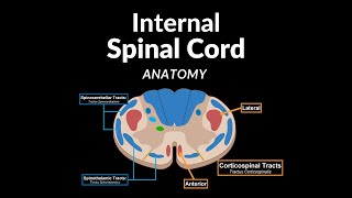
2 is nucleus gracilis and 3 is nucleus cuneatus
Amazing
Sooooooo gooodddddd
Why decussation occurs?
What's its significance?
Please tell the answer!
i suppose this is the lower medulla... as we are able to see the nucleus cuneatus and gracillis ..am i right ?
Can i get this table pic individually
What happens if medulla oblongata is constricted?
سبحان الله العظيم
Thank u
guys i dont understand the kind of fibres that goes to the gracilis nucleus,i understand that there coming from the lower part of the body but what is propreception what kind of fibres are they ???
They mainly come from muscle tendons and joints; so they help your brain understand the position of your joints and the stretch of the muscles :)
@@TaimTalksMed thanks a lot brother
Can I translate it into Uzbek and post it on the channel?
Absolutely! As long as you refer it back to the original video :)
Here after Waterboy 😂
wwooooowww👏👏👏👏👏
Where can I find this brain model?
I use the program Complete Anatomy :)
Blessings in an abundance of the faith. I salute you. 😘🙏Grace and peace be unto you and to this place. God bless you. 🤍🌈🤍Thank you. 💌Will you believe and receive Jesus Christ as your LORD and personal saviour??? I do. 😊
❤❤❤❤❤
Can i get these notes