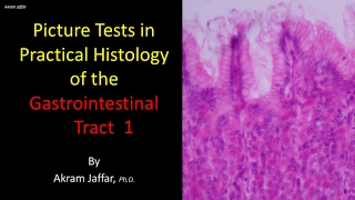
Picture tests in histology of the gastrointestinal system 3

Вставка
- Опубліковано 10 вер 2024
- After completion of this video you will be able to:
Differentiate between:
Endothelial cells lining haptic sinusoids and Kupfer cells
Orientations of muscle fibers in the muscularis externa.
Adventitia and serosa.
Acini of sero-mucous glands and their ducts.
Describe the structure of a hepatic lobule and the orientation of hepatic cords.
Identify:
Types of epithelium lining the gut: stratified squamous, simple columnar and describe their functions.
Glands associated with stomach, small and large intestine.
Layers of the gastrointestinal tube: mucosa, submucosa, muscularis, and adventitia.
Layers of mucosa: epithelium, lamina propria and muscularis mucosea.
Gastro-esophageal junction.
Sections of esophagus, stomach, small intestine, large intestine, and liver.
Gastric pits, parietal cells, mesothelial cells, hepatocytes, endothelial cells, and Kupfer cells
Presented and edited by Dr. Akram Jaffar, Ph.D.
This video and its channel are supported by "Human Anatomy Education" Page on Facebook / anatomyeducation
Subscribe to the channel to receive updates.
Feedback is highly appreciated from channel viewers
Some images, with gratitude, were cited in:
www.siumed.edu/...
www.drugdevelop...
www.doctorc.net...
www.histology.l...


Thank you so much! These series of videos have helped a lot.
You're amazing. Love your vids!
thanks so much, really helpfull, learned a lot
You're very welcome!
Great! Thank you!
You are welcome!
How to tell the gastroesophageal junction apart from intestinal metaplasia in Barret esophagus ?