අපි ECG කියවමු.. 1 කොටස How to read ECG basic rhythm in sinhala

Вставка
- Опубліковано 9 вер 2024
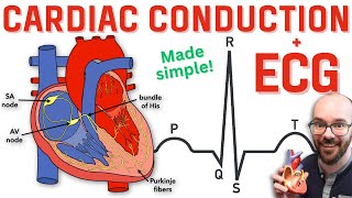
- A basic ECG rhythm, also known as normal sinus rhythm, represents the standard electrical activity of a healthy heart. Here's a detailed description of the key components of a basic ECG rhythm:
Normal Sinus Rhythm (NSR):
Origin: Originates from the sinoatrial (SA) node, the natural pacemaker of the heart, which is located in the right atrium.
Rate: The normal heart rate typically falls between 60 and 100 beats per minute in adults.
Steady Rhythm: Regular and consistent timing between heartbeats.
P Wave:
Description: The P wave is the first upward deflection on the ECG.
Significance: Represents atrial depolarization, indicating the contraction of the atria.
Normal Duration: Usually less than 0.12 seconds.
PR Interval:
Definition: Measured from the beginning of the P wave to the beginning of the QRS complex.
Significance: Represents the time taken for the electrical impulse to travel from the atria to the ventricles.
Normal Duration: Typically between 0.12 and 0.20 seconds.
QRS Complex:
Description: The QRS complex is the combination of the Q, R, and S waves.
Significance: Represents ventricular depolarization, indicating the contraction of the ventricles.
Normal Duration: Usually less than 0.12 seconds.
QT Interval:
Definition: Measured from the beginning of the QRS complex to the end of the T wave.
Significance: Represents the total time for ventricular depolarization and repolarization.
Normal Duration: Varies with heart rate; a corrected QT (QTc) is often used for comparison.
T Wave:
Description: The T wave is the upward deflection following the QRS complex.
Significance: Represents ventricular repolarization, indicating the relaxation of the ventricles.
Normal Characteristics: Usually in the same direction as the QRS complex but smaller.
In summary, a basic ECG rhythm, or normal sinus rhythm, is characterized by a regular heartbeat originating from the SA node, with distinct P waves, QRS complexes, and T waves. The intervals between these components fall within normal ranges. Any deviations from this pattern may indicate underlying cardiac issues, and a healthcare professional should interpret and analyze the ECG results for a comprehensive assessment of heart
ecg,ekg,ecg interpretation,ekg interpretation,ecg interpretation made easy,nursing school,nursing


Super, thanks
Great explanation doctor.
super sir. thank you so much. i am watching this after asking blame from by my consultant because not knowing how to interpret ecg. so this is very valuable for us sir
😊😊😊 you are welcome
Ammoooo...hina unaa..igenath gatta..the best teacher ❤
Thank you
Great explanation thank you
You are welcome!
මරු ❤
Docor u explained very wel..thank you very much
You are welcome
Great
Thank you
Please can you do physics and measurement topics
Good explanation
Can you do ECG lectures in English
Thanks you
English please
Good job. All the best Dinusha.
Thank you machan
sir i work in the private biomedical field , sir great explanation , let me more correct you v1 , v2 placement it should be 4 th intercostal line , thank you, sir please talk about arrithmia conditions and reasons , anti arrythmic drugs , their ecg changes and explanations to those changes , also talk about right side ecg placement and patterns , ..............etc
Thank you waruna
Thank you very much ayye🙂🙂🙂
❤❤❤❤❤
Thank you❤
Super❤️❤️❤️
Thank you
❤️❤️
🙂🙂
💯👌
Ube CCTV eka monapeththatada harawala thibune
Dyyo inna thannam waradiii . Incorrect chest leads placement.
😁 thank you doctor
අාාා 😟😛😛😛😛
ශෝක් නේ ,
හිනාවුනා ,
එතකොට අනිත් ටික කියලා දෙන්නේ කවුද ?
So do RS and CVS too.
දෙවැනි කොටසෙන්😂😂😂🙂🙂🙂🤗🤗
Ok i will do 😊😊🤗
Super, thanks