Cerebrum : Usmle Gross Anatomy || Relations and External features - Neuroanatomy animations

Вставка
- Опубліковано 11 лип 2024
- 📌 𝐅𝐨𝐥𝐥𝐨𝐰 𝐨𝐧 𝐈𝐧𝐬𝐭𝐚𝐠𝐫𝐚𝐦:- / drgbhanuprakash
📌𝗝𝗼𝗶𝗻 𝗢𝘂𝗿 𝗧𝗲𝗹𝗲𝗴𝗿𝗮𝗺 𝗖𝗵𝗮𝗻𝗻𝗲𝗹 𝗛𝗲𝗿𝗲:- t.me/bhanuprakashdr
📌𝗦𝘂𝗯𝘀𝗰𝗿𝗶𝗯𝗲 𝗧𝗼 𝗠𝘆 𝗠𝗮𝗶𝗹𝗶𝗻𝗴 𝗟𝗶𝘀𝘁:- linktr.ee/DrGBhanuprakash
Cerebrum: Gross anatomy, Relations and External features - Neuroanatomy animations
The cerebrum is the largest part of the brain, located superiorly and anteriorly in relation to the brainstem. It consists of two cerebral hemispheres (left and right), separated by the falx cerebri of the dura mater. Embryologically, the cerebrum is derived from the prosencephalon.
Anatomical Position and Structure
--------------------------------------------------------
The cerebrum is located within the bony cranium. It extends from the frontal bone anteriorly to the occipital bone posteriorly. Within the skull, the cerebrum fills the anterior and middle cranial fossae and is located above the tentorium cerebelli anteroposteriorly.
Internal Structure
-----------------------------
The cerebrum is comprised of two different types of tissue - grey matter and white matter:
Grey matter forms the surface of each cerebral hemisphere (known as the cerebral cortex), and is associated with processing and cognition.
White matter forms the bulk of the deeper parts of the brain. It consists of glial cells and myelinated axons that connect the various grey matter areas.
External Structure
------------------------------
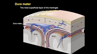
Externally, the cerebrum has a highly convoluted appearance, consisting of sulci (grooves or depressions) and gyri (ridges or elevations). It is divided into two anatomically symmetrical hemispheres by the longitudinal fissure - a major sulcus that runs in the median sagittal plane. The falx cerebri (a fold of dura mater) descends vertically to fill this fissure. The two cerebral hemispheres are connected by a white matter structure, called the corpus callosum.
The main sulci are:
------------------------------
Central sulcus - groove separating the frontal and parietal lobes.
Lateral sulcus - groove separating the frontal and parietal lobes from the temporal lobe.
Lunate sulcus - groove located in the occipital cortex.
The main gyri are:
----------------------------
Precentral gyrus - ridge directly anterior to central sulcus, location of primary motor cortex.
Postcentral gyrus - ridge directly posterior to central sulcus, location of primary somatosensory cortex.
Superior temporal gyrus - ridge located inferior to lateral sulcus, responsible for the reception and processing of sound.
Lobes of the Cerebrum
--------------------------------------
The cerebral cortex is classified into four lobes, according to the name of the corresponding cranial bone that approximately overlies each part. Each lobe contains various cortical association areas - where information from different modalities are collated for processing. Together, these areas function to give us a meaningful perceptual interpretation and experience of our surrounding environment.
Frontal Lobe
---------------------
The frontal lobe is located beneath the frontal bone of the calvaria and is the most anterior region of the cerebrum. It is separated from the parietal lobe posteriorly by the central sulcus and from the temporal lobe inferoposteriorly by the lateral sulcus.
The association areas of the frontal lobe are responsible for: higher intellect, personality, mood, social conduct, and language (dominant hemisphere side only).
Parietal Lobe
---------------------
The parietal lobe is found below the parietal bone of the calvaria, between the frontal lobe anteriorly and the occipital lobe posteriorly, from which it is separated by the central sulcus and parieto-occipital sulcus, respectively. It sits superiorly in relation to the temporal lobe, being separated by the lateral sulcus.
Its cortical association areas contribute to the control of: language and calculation on the dominant hemisphere side, and visuospatial functions (e.g. 2-point discrimination) on the non-dominant hemisphere side.
Temporal Lobe
-------------------------
The temporal lobe sits beneath the temporal bone of the calvaria, inferior to the frontal and parietal lobes, from which it is separated by the lateral sulcus.
The cortical association areas of the temporal lobe are accountable for memory and language - this includes hearing as it is the location of the primary auditory cortex.
Occipital Lobe
------------------------
The occipital lobe is the most posterior part of the cerebrum situated below the occipital bone of the calvaria. Its inferior aspect rests upon the tentorium cerebelli, which segregates the cerebrum from the cerebellum. The parieto-occipital sulcus separates the occipital lobe from the parietal and temporal lobes anteriorly.
#cerebrum #cerebrumanatomy #cerebrumanimation #neuroanatomy #cerebrumlecture #usmle #nationalexittest #fmge #mbbs #neuroanatomyvideos #neuroanatomylectures #medicalstudents #usmleprep #usmlepreparation #drgbhanuprakash #mbbslectures #usmle


📌 𝐅𝐨𝐥𝐥𝐨𝐰 𝐨𝐧 𝐈𝐧𝐬𝐭𝐚𝐠𝐫𝐚𝐦:- instagram.com/drgbhanuprakash
📌𝗝𝗼𝗶𝗻 𝗢𝘂𝗿 𝗧𝗲𝗹𝗲𝗴𝗿𝗮𝗺 𝗖𝗵𝗮𝗻𝗻𝗲𝗹 𝗛𝗲𝗿𝗲:- t.me/bhanuprakashdr
📌𝗦𝘂𝗯𝘀𝗰𝗿𝗶𝗯𝗲 𝗧𝗼 𝗠𝘆 𝗠𝗮𝗶𝗹𝗶𝗻𝗴 𝗟𝗶𝘀𝘁:- linktr.ee/DrGBhanuprakash
Is anatomy that simple 😳🙄.... you’re the best who makes anatomy simple and interesting 💝🤗👍
thank u so much
Sir thank you so much 🥲 today's is my exam
SO GOOD! WHAT THE BEST WAY EXPLAINING THIS CONFUSING TOPIC :D THIS WAY MAKES ME MORE UNDERSTAND ABOUT THEM.
Magician of anatomy ❤️🙏🙏🙏
Best comment I love it... thank you so much
Excellent work. You are a very good teacher. I was struggling with the topic until I came here, keep up the good work sir.
Thank you, I will
Thank you sir for great explanation🙏🙏
Thank You so much. Your videos are great
Mad respect, sir. You make the most complex items so easy.
Glad you like them!
Thank you so much sir ❤️
Neuroanatomy is ❤️
Ur most welcome
Thank you dr
So anatomy is beautiful
Extremely perfect, the method of illustration is superb, good luck
Thank you so much 😀
Thank you sir...
It is helping allot ✌️🤗
Ur most welcome
Thanks a lot for this amazing tutorial
Tysm
Thank you sir 🔥
Thank you sir
well explained
Extremely helpful ❤️
Tysm
Excellent video,thank you sir
Glad you liked it
It was very helpful sir thank you.
Looking for more😻😻😻
Tysm
Thanks sir,well explain
Ur most welcome
Very effective video,but there should be a detailed one.loved it
Ok next time
I love this person what a legend brother ❣️
Tysm
Hello
Fida hu tumpe meri ja bepanah
excellent lecture.
tysm
Nice explaination doctor 👌🏻👌🏻
Tysm
Great explanation
Tysm
Greaaaat ❤❤❤
tq sir
Sir pls explain about lymphatic system and endocrinology..
Thanks ✌🏻
Duhh thanx alot
I was just trapped in the same topic from last month and still it was so so confusing. Thanks again.....
Welcome 🤝🤝🤝
That cerebrum looks beautiful✨❤😍
thank you
Nice 💜💜💜
Thanks🥰
You’re welcome 😊
Thank you very much sir I always enjoy how you take time to explain anatomy
Sir. Your video is very nice but please increase the size of written headings or writing material ❣️
Sure, Noted
Sir how can we get a complete lecture of neuroanatomy
Sir. Is it possible to increase height by activating pituitary growth hormone after the growth plate is closure and How much inches grow possible?..
Thanks sir and cerebellum ?
Will upload soon
Make a video on mrcp and plab sil vous plait sir
sure
How can we cure the cerebellum shrinks?
Zhealthperformance
Sir aap hindi me bhi videos banaya kijiye na
i cant speak hindi well yaar thats the problem
Sir insula give me information because my brain not feel any emotions ?
Sir I want all the video of neuro anatomy.how can I get?😐
🔫
Sir. Is it possible to increase height by activating pituitary growth hormone after the growth plate is closure and How much inches grow possible?..
Sir. Is it possible to increase height by activating pituitary growth hormone after the growth plate is closure and How much inches grow possible?..