Answering a question we sometimes encounter from radiology residents, referring providers, and laypersons… The 4 main lung nodule margin categories are: • SMOOTH: margin with no perceptible projections, bulges, lumps, or indentations. Although most lung nodules with smooth margins are benign, 20% of malignant nodules have smooth margins & most metastatic nodules are smooth. • LOBULATED: at least one abrupt bulge in contour. Medium-high suspicion for malignancy. • SPICULATED: radial & unbranched thin linear opacities extend orthogonally from nodule margin into surrounding lung parenchyma, without reaching pleural surface. Very high suspicion for malignancy. • IRREGULAR: margin that isn’t smooth, lobulated, or spiculated. Indeterminate for benign vs. malignant. It can sometimes be challenging to distinguish whether a lung nodule margin is spiculated or irregular. Chest radiologists who specialize in lung cancer and other lung diseases (and publish a lot of the literature on lung nodules, their imaging, and outcomes) tend to be pretty rigorous and seasoned when making this distinction, and generally make this distinction with lower inter-reader variability. In practices where the distinction between “spiculated” and “irregular” is handled more liberally however, the predictive value of the term “spiculated” for malignancy may sometimes, as one might expect, differ from what is reported in medical research literature, published in textbooks, and traditionally taught in radiology residency.
I have had a history of benign lung nodules I. Recently, i had a preoperative screening CT scan and they found a new 15 mm nodule left lower lobe. The radiologist report says favor inflammatory or infectious process. I have had many bouts of pneumonia and flu. I also have had sepsis once. I also suffer from rheumatoid arthritis. I have never smoked. I have no family history of lung cancer. A CT scan or pet scan was recommended in 3 months. I'm truly nervous about the size of this one. Your video has helped me to understand very much. I'm hoping I'm okay
@JoyYacobacci-tx4xu I will pray for you. I understand your fear. I pray for both of us to have that peace that passes all understanding. I see the doctor next week for mine. Hopefully I will have some information. God bless you.
I wish there was a standard for radiology reports that included your standards listed here. I have yet to see such well written informed imaging studies in my health history that is packed with fragmented substandard reports. Good imaging technologists are 💎💫🌟 I hate it when a report doesn't inform. My last one had two lines of text. Lung nodules. No specifics regarding size, count, shape, content, characteristics. Patients often spend weeks gridlocked in insurance imposed hoops then wait weeks for an appointment. One study took eight months for an appointment!
I had a CT and shows I have a 2mm lower left lobe nodule. I'm 39 and vape heavily. Since April 2024 I've had bronchitis twice and pneumonia. August 2024 CT showed lungs were clear but found 2mm nodule. I've only had this cough for 4 months and actually vaping less in that time. But my cough won't go away. Do lung nodules make you cough? I'm waiting for appointment with respiratory specialist.
I had a small nodule removed during heart surgery small wedge it turned out to be adnocarcinoma why arent my nodules 7ml not being looked it went from 4 to 7 i just do not understand
I had RCC T1A Grade 2 at age 37. They just discovered a nodule on X Ray on the upper lobe of my left lung. Going in for a CT tomorrow and I am hopeful it’s not what it could be. I am confused about the risk to patients who have been NED for years vs patients with active disease.
Had influenza A in January that then became atypical pneumonia. As of April, still had an LUL GGO possibly 22mm, and concerningly to me, a new 6mm nodule with four less-than 4mm nodules in LLL. I live in North Central WI which is an endemic area for Blastomycosis. Next CT in beginning of July. If nodules are larger FDG-PET is next and possibly robtic brochoscopy & sputum cytology for aspergilliosis & blastomycosis. Very worried I have Adenocarcinoma because I'm an 80pk-yr smoker
I have PsA and developed shortness of breath and low oxygen sats so was referred to Pulmonology. A high-resolution CT showed findings consistent with bronchiolitis obliterans. Incidentally noted was a 5-mm solid nodule. A followup CT 1 year later showed the nodule was down to 3 mm, so I was relieved. However, a couple months later, the report was amended to show the nodule was still 5 mm. I am listed as high risk for malignancy because of my use of biologics and I'm assuming my age of 68. I will be having another high-resolution CT in May. My question is can a nodule remain stable for a few years but then go on to become a cancer?
Thank you very much for your videos! Could you please tell me where the information about calcification patterns comes from? In our country, amorphous calcification is considered to be a benign sign.
This approach to lung nodule calcification patterns was taught to us when I was resident, and continues to be what my colleagues and I continue to teach our residents today. It appears in textbooks we assign for reading, and in the scientific literature too. Take for example, the chest radiology textbook "Diagnostic Thoracic Imaging" by Wallace T. Miller (a radiology textbook favored at Penn): "Amorphous, irregular, punctate, and eccentric patterns of calcification have been identified in a variety of malignancies including bronchogenic carcinoma, carcinoid tumors, and metastasis." "It must be remembered that calcification alone is not diagnostic of a granuloma or a benign condition. Calcification will be present in 6% to 14% of primary lung carcinomas. However, the calcification in cancer is typically amorphous or stippled in character, different than the patterns of calcification which are specific for granulomas." Similar discussions of the different lung nodule calcification patterns and their implications go back for decades in the scientific literature. Take for example the 1993 AJR paper "CT of the Lung: Patterns of Calcification and Other High-Attenuation Abnormalities" by Chai and Patz: "Approximately 6% of all primary lung cancers show a punctate, amorphous, or reticular pattern of calcification on CT scans. This variation is probably due to several different causes: (1) engulfment of benign calcification by the tumor as is seen in scar carcinoma, (2) dystrophic calcification arising from necrosis within the tumor, and (3) calcium deposition resulting from secretions by the tumor."
Can you create a video explaining lung nodules and their risk of malignancy for patients? I have 8 solid lung nodules, all measuring 6mm and smaller, located in the right lung. Stable on 6 month’s scan. One of these nodules is 6mm in size and spiculated. When I researched spiculated nodules, I learned that they are almost always malignant, with a predictive value of up to 90%. I also came across your comment on here stating that even small spiculated nodules are almost always malignant. So, regardless of their small size, spiculated nodules seem to consistently indicate malignancy. I'm confused as to why doctors still recommend a watch and wait approach when spiculation is such a certain indicator of malignancy. My doctor couldn't provide me with clear answers. He did mention that spiculation, along with other nodules, can sometimes be caused by infection or inflammation. This conflicting information is making it difficult to make a decision. I don't want to hold onto false hope. Should I take a more proactive approach in my case?
Pray and eat things that fight cancer and parasite. I have one in my lung and possibly neck. I'm praying eating garlic, vitamin c, spinach, turmeric powder.
I have a 6mm nodule is my posterior basilar lower right lobe but it doesn’t say anything about it being anywhere near the pleura. I’m supposed to go for CT scans every 6-12 mos for a bit to monitor it.
Have a nodule one is 8mil an no body told me had covig 2020 an alot colds copd quick patches are not cover under my insurance nicotine patch does increase chance it will go it round tail on it radiation therapy target area an schink it what chanes of me didnt want biosy want secons opinion on it
Ct scan showed 5 solid lung nodules between 4-5mm in size 2 are calcified granuloma, 2 are perifisual 1 nodule is Spiculated. After doing some research, it seems most likely that the Spiculated nodule is malignant. how often do you have cases of Spiculated lung nodules that turn out to be benign? Do such cases occur often or are they really that rare?
My colleagues and I tend to use the term “spiculated” in nodules over 1 cm, as it’s pretty tough to distinguish between spiculated margins vs irregular margins when a nodule is under 1 cm. In lung nodules that we *do* call spiculated, the large majority are malignant. The few cases where the nodule ended up being benign were generally those that appeared in the background of severe emphysema, which can oftenmake infectious opacities look overly suspicious, since there’s not much parenchyma left and consolidation has to follow the architecture of whatever is left. Hope that helps!
My ct scan with contrast stated findings of stable 3mm lower right lower lobe . They did the scan because I had viral covid pneumonia. My question is I’m wonder why they used that word stable ? Ty
The radiologist who interpreted your CT scan probably noticed that the 3 mm right lower lobe lung nodule was also visible on an older CT and hadn't changed.
I have a lower right pulmonary lung nodule. In march of 2018 it was 2mm and now it’s 9mm……. Its round. I go for follow up CT scan next month. Is this big or small? Does growth automatically mean cancer? I’m 40 and smoked throughout my 20’s
Full chest LDCT only showed that 1 9mm nodule. They are going to take it out in January. They said based on location, size, slow doubling time and well-circumscribed, smooth and round it has a low chance of being malignant. Based on where its located they cant biopsy. So they are going to wedge resection.
I had 5 days of prednisone for condrocitis no cough never sick just chest pain in ribs from breast bone going around to back painful to touch so after 5 days I wasn't satisfied so I went got a x-ray they say I have a nodule gave me doxycycline for two weeks still no symptoms other than tenderness want me to comeback 2 weeks for another X-ray I have drank mineral oil in the past not sure if it matters I'm 42 have had two dvt and been in remission of a very rare disease since 2006 called multicentric castlemans disease treated with chop and rituxin I'm scared to death they said it was a nodule not a mass but if seen in a xray must had been decent size
That is correct, the target audience for the talks in this channel are physicians and physicians-in-training. However, if you notice topics in this channel that might be ones that may interest many laypersons/patients, let me know and we can build versions for non-medical professionals too.


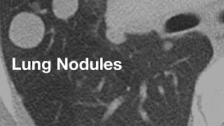


Answering a question we sometimes encounter from radiology residents, referring providers, and laypersons…
The 4 main lung nodule margin categories are:
• SMOOTH: margin with no perceptible projections, bulges, lumps, or indentations. Although most lung nodules with smooth margins are benign, 20% of malignant nodules have smooth margins & most metastatic nodules are smooth.
• LOBULATED: at least one abrupt bulge in contour. Medium-high suspicion for malignancy.
• SPICULATED: radial & unbranched thin linear opacities extend orthogonally from nodule margin into surrounding lung parenchyma, without reaching pleural surface. Very high suspicion for malignancy.
• IRREGULAR: margin that isn’t smooth, lobulated, or spiculated. Indeterminate for benign vs. malignant.
It can sometimes be challenging to distinguish whether a lung nodule margin is spiculated or irregular. Chest radiologists who specialize in lung cancer and other lung diseases (and publish a lot of the literature on lung nodules, their imaging, and outcomes) tend to be pretty rigorous and seasoned when making this distinction, and generally make this distinction with lower inter-reader variability.
In practices where the distinction between “spiculated” and “irregular” is handled more liberally however, the predictive value of the term “spiculated” for malignancy may sometimes, as one might expect, differ from what is reported in medical research literature, published in textbooks, and traditionally taught in radiology residency.
2nd year Rad res and this was such a concise high yield review. Thank you!
Thanks for the kind feedback!
I have had a history of benign lung nodules I. Recently, i had a preoperative screening CT scan and they found a new 15 mm nodule left lower lobe. The radiologist report says favor inflammatory or infectious process. I have had many bouts of pneumonia and flu. I also have had sepsis once. I also suffer from rheumatoid arthritis. I have never smoked. I have no family history of lung cancer. A CT scan or pet scan was recommended in 3 months. I'm truly nervous about the size of this one. Your video has helped me to understand very much. I'm hoping I'm okay
Prayers too you 🙏. They just found one 1 an half cm.on my left ..I'm scared😭
@JoyYacobacci-tx4xu I will pray for you. I understand your fear. I pray for both of us to have that peace that passes all understanding. I see the doctor next week for mine. Hopefully I will have some information. God bless you.
@Peaceful-hi9iy Prayers too you also an God Bless 🙌
@@Peaceful-hi9iy pray for me and every soul too.
@meldj9352 I am praying for you right now. God will take care of you and he loves you very much. Me too, in Jesus.💖❤️❤️
I wish there was a standard for radiology reports that included your standards listed here. I have yet to see such well written informed imaging studies in my health history that is packed with fragmented substandard reports. Good imaging technologists are 💎💫🌟
I hate it when a report doesn't inform. My last one had two lines of text. Lung nodules. No specifics regarding size, count, shape, content, characteristics. Patients often spend weeks gridlocked in insurance imposed hoops then wait weeks for an appointment. One study took eight months for an appointment!
I had a CT and shows I have a 2mm lower left lobe nodule. I'm 39 and vape heavily.
Since April 2024 I've had bronchitis twice and pneumonia.
August 2024 CT showed lungs were clear but found 2mm nodule.
I've only had this cough for 4 months and actually vaping less in that time.
But my cough won't go away.
Do lung nodules make you cough?
I'm waiting for appointment with respiratory specialist.
I had a small nodule removed during heart surgery small wedge it turned out to be adnocarcinoma why arent my nodules 7ml not being looked it went from 4 to 7 i just do not understand
Highly usesul presentation, thank you sir 👏👏👏👏
You are most welcome!
I love this video! It will be super helpful for my research to look into the lung mets. Thanks a lot :)
Great presentation
I had RCC T1A Grade 2 at age 37. They just discovered a nodule on X Ray on the upper lobe of my left lung. Going in for a CT tomorrow and I am hopeful it’s not what it could be. I am confused about the risk to patients who have been NED for years vs patients with active disease.
Had influenza A in January that then became atypical pneumonia. As of April, still had an LUL GGO possibly 22mm, and concerningly to me, a new 6mm nodule with four less-than 4mm nodules in LLL. I live in North Central WI which is an endemic area for Blastomycosis. Next CT in beginning of July. If nodules are larger FDG-PET is next and possibly robtic brochoscopy & sputum cytology for aspergilliosis & blastomycosis. Very worried I have Adenocarcinoma because I'm an 80pk-yr smoker
I have PsA and developed shortness of breath and low oxygen sats so was referred to Pulmonology. A high-resolution CT showed findings consistent with bronchiolitis obliterans. Incidentally noted was a 5-mm solid nodule. A followup CT 1 year later showed the nodule was down to 3 mm, so I was relieved. However, a couple months later, the report was amended to show the nodule was still 5 mm. I am listed as high risk for malignancy because of my use of biologics and I'm assuming my age of 68. I will be having another high-resolution CT in May. My question is can a nodule remain stable for a few years but then go on to become a cancer?
Interesting question, let me know if you receive an answer
@@joejudy1 Since you replied to my comment, if I get an answer you should be alerted, but I'll let you know as well. 👍
Thank you very much for your videos! Could you please tell me where the information about calcification patterns comes from? In our country, amorphous calcification is considered to be a benign sign.
This approach to lung nodule calcification patterns was taught to us when I was resident, and continues to be what my colleagues and I continue to teach our residents today. It appears in textbooks we assign for reading, and in the scientific literature too.
Take for example, the chest radiology textbook "Diagnostic Thoracic Imaging" by Wallace T. Miller (a radiology textbook favored at Penn):
"Amorphous, irregular, punctate, and eccentric patterns of calcification have been identified in a variety of malignancies including bronchogenic carcinoma, carcinoid tumors, and metastasis."
"It must be remembered that calcification alone is not diagnostic of a granuloma or a benign condition. Calcification will be present in 6% to 14% of primary lung carcinomas. However, the calcification in cancer is typically amorphous or stippled in character, different than the patterns of calcification which are specific for granulomas."
Similar discussions of the different lung nodule calcification patterns and their implications go back for decades in the scientific literature. Take for example the 1993 AJR paper "CT of the Lung: Patterns of Calcification and Other High-Attenuation Abnormalities" by Chai and Patz:
"Approximately 6% of all primary lung cancers show a punctate, amorphous, or reticular pattern of calcification on CT scans. This variation is probably due to several different causes: (1) engulfment of benign calcification by the tumor as is seen in scar carcinoma, (2) dystrophic calcification arising from necrosis within the tumor, and (3) calcium deposition resulting from secretions by the tumor."
Can you create a video explaining lung nodules and their risk of malignancy for patients? I have 8 solid lung nodules, all measuring 6mm and smaller, located in the right lung. Stable on 6 month’s scan. One of these nodules is 6mm in size and spiculated. When I researched spiculated nodules, I learned that they are almost always malignant, with a predictive value of up to 90%. I also came across your comment on here stating that even small spiculated nodules are almost always malignant. So, regardless of their small size, spiculated nodules seem to consistently indicate malignancy.
I'm confused as to why doctors still recommend a watch and wait approach when spiculation is such a certain indicator of malignancy. My doctor couldn't provide me with clear answers. He did mention that spiculation, along with other nodules, can sometimes be caused by infection or inflammation. This conflicting information is making it difficult to make a decision. I don't want to hold onto false hope. Should I take a more proactive approach in my case?
How r u feeling now
you much go for two test Bronchsopy & Pet/Ct Scan. then you find out about true. what happen in your body.
@@michaelliangliang4677PET CT definitely.
Pray and eat things that fight cancer and parasite. I have one in my lung and possibly neck. I'm praying eating garlic, vitamin c, spinach, turmeric powder.
I have a 6mm nodule is my posterior basilar lower right lobe but it doesn’t say anything about it being anywhere near the pleura. I’m supposed to go for CT scans every 6-12 mos for a bit to monitor it.
Have a nodule one is 8mil an no body told me had covig 2020 an alot colds copd quick patches are not cover under my insurance nicotine patch does increase chance it will go it round tail on it radiation therapy target area an schink it what chanes of me didnt want biosy want secons opinion on it
Ct scan showed 5 solid lung nodules between 4-5mm in size
2 are calcified granuloma, 2 are perifisual
1 nodule is Spiculated.
After doing some research, it seems most likely that the Spiculated nodule is malignant.
how often do you have cases of Spiculated lung nodules that turn out to be benign? Do such cases occur often or are they really that rare?
My colleagues and I tend to use the term “spiculated” in nodules over 1 cm, as it’s pretty tough to distinguish between spiculated margins vs irregular margins when a nodule is under 1 cm. In lung nodules that we *do* call spiculated, the large majority are malignant. The few cases where the nodule ended up being benign were generally those that appeared in the background of severe emphysema, which can oftenmake infectious opacities look overly suspicious, since there’s not much parenchyma left and consolidation has to follow the architecture of whatever is left. Hope that helps!
What does non-calcified nodule mean?
My ct scan with contrast stated findings of stable 3mm lower right lower lobe . They did the scan because I had viral covid pneumonia. My question is I’m wonder why they used that word stable ? Ty
The radiologist who interpreted your CT scan probably noticed that the 3 mm right lower lobe lung nodule was also visible on an older CT and hadn't changed.
also, calcified modules are not of concern
I have a lower right pulmonary lung nodule. In march of 2018 it was 2mm and now it’s 9mm……. Its round. I go for follow up CT scan next month. Is this big or small? Does growth automatically mean cancer? I’m 40 and smoked throughout my 20’s
What did you find out?
Full chest LDCT only showed that 1 9mm nodule. They are going to take it out in January. They said based on location, size, slow doubling time and well-circumscribed, smooth and round it has a low chance of being malignant. Based on where its located they cant biopsy. So they are going to wedge resection.
@@brianwillow4898 That's good,best of luck with surgery,I have one that's 3mm I saw it on my med records about two years ago,no one even told me.
@nikolainewton2295 You should been told about it even though doctor dismissed because of size.
😊@@nikolainewton2295
Excellent. Thanks.
I had 5 days of prednisone for condrocitis no cough never sick just chest pain in ribs from breast bone going around to back painful to touch so after 5 days I wasn't satisfied so I went got a x-ray they say I have a nodule gave me doxycycline for two weeks still no symptoms other than tenderness want me to comeback 2 weeks for another X-ray I have drank mineral oil in the past not sure if it matters I'm 42 have had two dvt and been in remission of a very rare disease since 2006 called multicentric castlemans disease treated with chop and rituxin I'm scared to death they said it was a nodule not a mass but if seen in a xray must had been decent size
👏👏👏
I'm not a doctor. This is not written for the masses just trying to figure out about basic lung nodule info.
That is correct, the target audience for the talks in this channel are physicians and physicians-in-training. However, if you notice topics in this channel that might be ones that may interest many laypersons/patients, let me know and we can build versions for non-medical professionals too.
Unfortunately, if you were recently diagnosed, this will all make perfect sense very soon.