I was taught that when looking for pathological Q waves, you look in the inferior leads mainly. The reason being that, leads v1 & v2 (septal) should be negative always so it's not reliable. Also lead III will appear to have a path. q wave that really isn't pathological, so you must find it in 2 or more contiguous leads.
Excellent, quality video. She is calm, confident in her delivery and precise. 100% perfect video. I have learnt so much by watching this video. Great Teacher! Thank you making this video!
Hey nice vid with clear explanations and diagrams. I thought it would've been perfect if you explained the electrophysiology behind why scar tissue results in a Q wave as no one seems to address this
@@Bobcheloo the reason is pretty simple the scar tissue ( which essentially is a dead tissue ) is creating an electrical window so when the depolarisation vector of other side is propagating, it gets detected by the electrical lead adjacent to scar tissue, for it the depolarisation vector should appear moving away, so the adjacent lead detects it as a downward deflection which essentially forms the deep Q wave.
i just started my clinicals , we were asked to look into the Q wave and i was blank. and this explained pretty much everything i need to know , thanks a lot :) and keep up the good work :)
I really enjoyed your lesson I'm a disabled veteran and I have a slight miss trust in the Dr.'s at my VA I have been complaining of heart issues since the 1990's , approx 6 years ago I ended up having a triple by pass and the surgeon who did this told me that this might have probably been avoided if the VA would have taken better precautions early on so I was thrilled to see instructions on this, I was a medic in the service not war time so I really did not get the chance to learn then
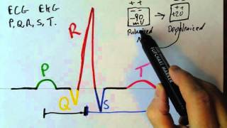
Lower case letters are used to denote a wave form that does not reach at least 3 small squares (3 mm) in the + or - from the isoelectric line, thus the above initial complex appears to be correctly written as: q R S
Hey, thank you for the explanation. I'm quite struggling to understand the graphic before. But I'm still wondering why the mi scar make the q wave more deeper. Can somebody explain it?
It creates a communication from inside the heart to the outer wall of the heart. What the ecg is showing from a DEEP Q wave is that we probably have a communication between the septum( inside the heart ) to the outside ( Basically if you put a electrode inside a ventricle, all you would see is a big Q wave, because the depolarization occurs from inside to the outside ). ( I'm a student, not a physician and I'm only saying what I think I read from the book I'm reading )
I could never understated these q waves earlier. Thank you. It would be greatly appreciated if a normal ECG and q waves pattern was shown side by side. If we know normal Ecg by heart, then we world pick up abnormalities in an instant ima!
I am studying for step now and your videos are helping me so much! I do wish you had the videos groups by subject so it was easier to find relevant ones. I don't know how you do that, but another channel I subscribe to has it set up like that.
Since in v1 to v3 the QRS is normaly negative, the R wave in v1 to v3 is not gonna be an upward spike, but a downward spike, so at 7:54 how can I be sure that it is not an R wave but a pathological Q wave?
Nevermind, I found out that the R wave in v1 to v3 is still upward, but just very small. Si if an R wave is absent, and after the P wave there isna downward spike, that is a Q wave.
Nice video, but I have a question about one of the interpretations. It, to me, appears to be a LBBB. But, i can't measure the width of the QRS since it's not a real ECG. I also see R waves in leads II and III. This is the ECG I'm referring to: i.imgur.com/jfP6Lwt.png


This really helped me understand the emphasis of looking for Q waves and why. Thank you.
I was taught that when looking for pathological Q waves, you look in the inferior leads mainly. The reason being that, leads v1 & v2 (septal) should be negative always so it's not reliable. Also lead III will appear to have a path. q wave that really isn't pathological, so you must find it in 2 or more contiguous leads.
i was looking for such a comment ,to be honest its not clear
Excellent, quality video. She is calm, confident in her delivery and precise. 100% perfect video. I have learnt so much by watching this video. Great Teacher! Thank you making this video!
Hey nice vid with clear explanations and diagrams. I thought it would've been perfect if you explained the electrophysiology behind why scar tissue results in a Q wave as no one seems to address this
do you know the answer to this? I have been looking everywhere for this
@@Bobcheloo the reason is pretty simple the scar tissue ( which essentially is a dead tissue ) is creating an electrical window so when the depolarisation vector of other side is propagating, it gets detected by the electrical lead adjacent to scar tissue, for it the depolarisation vector should appear moving away, so the adjacent lead detects it as a downward deflection which essentially forms the deep Q wave.
i just started my clinicals , we were asked to look into the Q wave and i was blank. and this explained pretty much everything i need to know , thanks a lot :) and keep up the good work :)
An absolutely brilliant lecture about pathologic Q,wave, many thanks Miss
This was fantastic, thank you. If I had any misunderstandings about a pathological Q-wave, I don't now.
Are u using zoom to describe? I liked your style. Thanks 🙏 for sharing.
very smart doctor explained pathological pwave axactly and clearly... a understood more than what i am not benifited from other expert doctors.. great
I really enjoyed your lesson I'm a disabled veteran and I have a slight miss trust in the Dr.'s at my VA I have been complaining of heart issues since the 1990's , approx 6 years ago I ended up having a triple by pass and the surgeon who did this told me that this might have probably been avoided if the VA would have taken better precautions early on so I was thrilled to see instructions on this, I was a medic in the service not war time so I really did not get the chance to learn then
Thank you for your service
Lower case letters are used to denote a wave form that does not reach at least 3 small squares (3 mm) in the + or - from the isoelectric line, thus the above initial complex appears to be correctly written as: q R S
I have never understood the significance of pathological q waves until now! Thank you!!!
Good learning video. You are a simple lady.
Your tutorial was really really great. Thank you so much. Now I understand Q waves. Please show more tutorials. Ok. You are a very good teacher.
Hey, thank you for the explanation. I'm quite struggling to understand the graphic before. But I'm still wondering why the mi scar make the q wave more deeper. Can somebody explain it?
It creates a communication from inside the heart to the outer wall of the heart. What the ecg is showing from a DEEP Q wave is that we probably have a communication between the septum( inside the heart ) to the outside ( Basically if you put a electrode inside a ventricle, all you would see is a big Q wave, because the depolarization occurs from inside to the outside ).
( I'm a student, not a physician and I'm only saying what I think I read from the book I'm reading )
finally i found this video! starting from the very basics was the best thing....thank you!!!
Doesn’t have the ECG at 11:46 at V2 and V3 pathologic Q wave and followed ST segment elevation?
Best video I’ve ever watched regarding EKG
I could never understated these q waves earlier. Thank you.
It would be greatly appreciated if a normal ECG and q waves pattern was shown side by side.
If we know normal Ecg by heart, then we world pick up abnormalities in an instant ima!
Excellent description
Excellent teaching. Thank you ❤❤❤❤❤❤
Thank you for your support.
Awesome explanation.
I really appreciate you’re work , today I clear my confusion about Q WAVE IN ECG
Super helpful and straight forward with many examples. Thank you!
Thank you so much It Is very interessting lesson.👍
Salam stay blessed u made Ecg understanding easy.Superb and Flawless.Thanks.
Thank you, this truly is extremely helpful for Q waves!
thank you for making this video
Nice explanation as well.
really great all ur videos.i always wait to see ur videos
The pathological q waves that you are pointing out, could be a normal qs deflection?
Great video, thanks!
Thank you for a very informative lesson.
Glad it was helpful!
Totally professional. Thanks for your time and teachings.
Excellent video and very helpful with all of the examples. Thank you so much!
Could you enlarge the EKG ? Really small.
good work....thanks
very clear explanations. God bless you.
It was a session of pure bliss
Awesome teacher 👍🏼🙏🏻
Thank you for your kind comment.
This helped me understand pathologic Q waves. Thanks!
By this videos I understood a lot then books ..thank u so much for such beautiful explanation .
Flawless explanation👏👏👏
We need more videos in ECG PLZZ
Great learning session, thank you
My pleasure!
Very helpful, much appreciated.
Can u plz explain 9:36 why in lead v3 (at bottom) u didn't consider st elevation..?
Really a helpful lecture.Plz go ahead.
This video is amazing! Thank you!
Thank, thank, thank! This cleared up some details I was muddled on. Great video, wonderful instruction!
I am studying for step now and your videos are helping me so much! I do wish you had the videos groups by subject so it was easier to find relevant ones. I don't know how you do that, but another channel I subscribe to has it set up like that.
This was amazing
Thanks, It is an important material, with a very nice and clear presentation! Good Luck with the future videos.
Excellent content , thank you 🙏
Can you interprete results that I have ecg results for Lead III and V6 abnormal q waves with poor r wave in V2
Thank you very much.
You have a sweet and very clear voice .
Eloquently explained into simplistic terms... thank you !!!
Informative video
Thank you so much.
Wonderful video! Great explaining and easy to follow;)!
Since in v1 to v3 the QRS is normaly negative, the R wave in v1 to v3 is not gonna be an upward spike, but a downward spike, so at 7:54 how can I be sure that it is not an R wave but a pathological Q wave?
Nevermind, I found out that the R wave in v1 to v3 is still upward, but just very small. Si if an R wave is absent, and after the P wave there isna downward spike, that is a Q wave.
WOUH, So well explained, so Easy made.
great . but in the last 2 ecg there t wave inversion . why it is like this
Where is part 2 ?
Please I need it
Nice video. Very informative and to the point.
Nice work
Excellent lecture ....good work!
Thank you.
Fantastic
Wow wonderful explanation ,thank you
one of the best videos ever !! 🤘
Thank you so much
Thank you very much you made ECG so easy for me
Thank you for simplifying. Thanks for doing this.
excellent video and explanation! thank you
Excellent presentation 😊❤
thank you - this was very helpful
Thank so much .
After watching your video I can confidently say I can recognise a pathologic Q wave:) very simple way of explanation :)
THANK YOU
Brilliant, thank you dear
This is a great informative lecture for me thanks alote mam
Very nice
You're a great teacher!!
Thank you!
Thank you for explaining it step by step
Good Video, thank you!
Very clearly explained. Thank you!
Very nice and very clear explanation thanks alot
12:55 there's st elevation in v1 to v4 right?
Super helpful thank you¡
This is great
Phenomenal video... thank you :)
awesome explanation! thank you so much!
Nice vid!
Really helpful.
Nice video, but I have a question about one of the interpretations. It, to me, appears to be a LBBB. But, i can't measure the width of the QRS since it's not a real ECG. I also see R waves in leads II and III. This is the ECG I'm referring to: i.imgur.com/jfP6Lwt.png
great
like the way of explaining, really easy.
Enjoyed your video. Thank you!