Thyroid Ultrasound Normal Vs Abnormal Image Appearances Comparison | Thyroid Pathologies USG

Вставка
- Опубліковано 1 сер 2024
- Thyroid Ultrasound Normal Vs Abnormal Image Appearances Comparison | Thyroid Pathologies USG
*Timestamps:
Intro: 0:00
Normal: 0:08
Thyromegaly: 1:00
Nodular Hyperplasia: 2:09
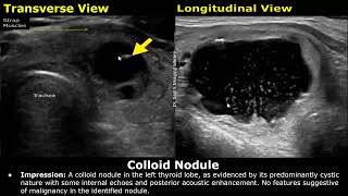
Colloid Nodule: 3:03
Follicular Adenoma: 3:57
Follicular Carcinoma: 4:39
Papillary Carcinoma : 5:22
Cystic Papillary Cancer: 6:11
Anaplastic Cancer: 6:42
Lymphoma: 7:08
Metastases: 7:38
Hashimoto's Thyroiditis: 7:56
Graves' Disease: 9:12
Subacute Thyroiditis: 10:19
Outro: 10:50
You can support our work with a donation:
Airtm: drsamslibrary@gmail.com
Patreon: patreon.com/drsamsimaginglibrary
Related: neck sonography, imaging, images, radiology, scan, scanning, goiter, hyperthyroidism, hypothyroidism, hypervascularity, longitudinal, transvers, cases, diseases, views, planes, grayscale, ultrasonography pathology, mets, region, enlargement, enlarged, doppler, hypervascularity, thyroid inferno, lesions, mass, masses hypoechoic, hyperechoic echogenic


Watch More Ultrasound Videos: ua-cam.com/play/PL4cRFWfjMmf_P02uIGRTFiYNozGKuILAX.html
Abdominal Ultrasound: ua-cam.com/play/PL4cRFWfjMmf_8Rxbgv4Ru77mxhAE9mN2L.html
Gynecological Ultrasound: ua-cam.com/play/PL4cRFWfjMmf-J4vhXPWZbx5uRIwq77EBd.html
Obstetric Ultrasound: ua-cam.com/play/PL4cRFWfjMmf-M-IH-Bq-LY-07LeEFdi5T.html
I subscribe your channel
@@irfanullah9581 Thank you for subscribing!
Please share thyroid ultrasound reporting
@@sanaaslam4262Yes it will be uploaded in the future.
Very good presentation, dr. Sam. Keep up the good work! All the best from Romania!
Thank you so much for watching!
Very good work Dr Sam (all your videos)! You are really helpful..all the best from Greece!
Most Welcome and thanks for watching! Greetings!
Thank you Dr Sam for this great lecture .
Most Welcome Sir!
Your videos are always the best and to the point
Thank you very much for watching!
Very informative ❤
Thanks Dr Sam for this nice lecture. We need more videos of ultrasound
Thank you very much!
that was so informative short and to the point. thanks
Most Welcome!
Nice explanations, many thanks!
Thank you so much for watching!
Thank you Dr. Sam 🙏
Most Welcome!
Thanks for those useful videos . Please keep making them 💐
Most Welcome and thanks for watching!
Great lecture, thank you Dr Sam
Most Welcome!
gracias por realizar estos videos desde República Dominicana me ha servido mucho para mis practicas de sonografia
Most Welcome and Thanks for watching!
Thank you Dr Sam.
Always welcome
Thank you for this. Keep it up sir.
Thank you so much for watching!
Thankyou for this.. Appreciate it😊
Most Welcome and thanks for watching!
Tanks a lot Dr Sam
Most Welcome!
Amazing job , God bless u sir .
Thank you very much
Very nice and informative. Thank you
Most Welcome!
Thanks Dr Sam for the lecturs
Most Welcome!
Thanks for sharing Dr
Your Welcome!
thanku dr sam. very informative short lecture. 👍
Most Welcome and thanks for watching!
Great one, thanks alot
Most Welcome!
Thank you
Sir really adore you i have learned so much from your videos God bless you sir
Thank you much for watching! Really appreciate it! God bless you too!
Wow!..very informative...God bless sir...
Thank you very much for watching!
Well done Dr Sam excellent demonstration.plz guide us about probing
Thanks for watching! You can watch this video regarding probe positioning: ua-cam.com/video/yGg7Ci88HrQ/v-deo.html
Love your vedios ❤
Excellent 👌
Thank you very much for watching!
Excellent session thanks a lot for favour sir
Most Welcome!
Thank you!
Your Welcome!
Excellent sir.
Such videos will help us Sir
Thank you very much!
U have very nicely define sonographic anatomy and pathology.
Thank you so much for watching!
GREAT SIR.... JUST GREAT VIDEO.
Thanks for watching!
Thank you I got photos but my doctor didn’t review them yet and I compared them using your video.
Most Welcome!
appreciate your efforts
Thank you very much!
You are educating us with nice presentation !! THANK you Sir 🙏🙏🙏
Thanks for watching!
Thank you very much. Very helpful. Greetings from Indonesian Sonographer
Most Welcome! Greetings!
Thanks for information
Most Welcome!
Thank u!
Your Welcome!
Thanks so much
Most Welcome!
Very Very thankful to Dr. Sam for you detail explanation. One suggestion that the translation on word coming in video get obscure the image and measurement you have shown, I think there is no need of translation in English . Otherwise all videos are excellent. Thank you
Thank you very much for watching! Really appreciate it! Yes, the translation and subtitles can obscure the image and the description. You can turn them off if needed.
Thanks Dr
Your Welcome
Excellent Sir
Thank you!
Thank you.
You're welcome!
Well done 👍...bro plz make a video about other lesions in the neck like ranula, brachial cyst, dermoid, ectopic thymus, ets..with detail thanx in advance
Thank you for watching and for your suggestions!
🥺🥺🥺great lectures sir ❤️
Thanks for watching!
Thank you sir
Most Welcome!
MARVELLOUS SIR.
Thanks for watching!
Sir, you are genius 💝
Thank you very much!
Thanks you very much Dr Sam. Good work. I want to know about the use of EU-TIRADS classification.
Most Welcome and thanks for your request!
Hello Dr Sam i was wondering if u can help me interpret what the radiologist reported on my neck ultrasound. My neck has been sore and swollen for the past 6 months now and I'm convinced I feel something hard near my thyroid which is causing both sides of my neck to swell. The radiologist report said I have two HYPOECHOIC nodules in my neck one. right one is 4mm. 7mm on the left lobe My right submandular nodes are 2.2cm by 0.8. Left 1.5cm by 0.7. is this within normal size range? I have a submental probable node that contains a SMALL ECHOGENIC FOCUS with no shadowing? Are hypoechoic nodules more likely to be cancerous? There is not nodules on my thyroid gland but there is nodules on my neck
Hello! Good day! It is difficult to accurately comment whether these nodules are cancerous or not based on an ultrasound report alone. It is best to consult with an ENT specialist because further investigation is required which includes clinical exam, lab tests as well as imaging findings. Best Wishes!
Thanks Dr. Sam! Can you make a video talking upper, mid, and lower poles for the thyroid for left and right lobe ?
Thanks for watching and thanks for your suggestion!
Thank you for useful and practical videos. I love your channel.
Are you agree with this point about hashimoto that colour Doppler study usually shows normal or decreased flow, but occasionally there might be hypervascularity?
Thank you very much for watching! Yes, I agree. It can have variable appearances in some cases
thank you or explaining with images .
Most Welcome and thanks for watching!
Thank you, Dr. Sam. I had an US early 2023 and it presented with the thyroid gland being atrophic in size and homogeneous. When I was 20 y.o I had RAI uptake, I had hyperthyroidism. It keeps going from hypo to hyper. Can you tell me more about the atrophic size and why. I still am having lots of symptoms and I am on Synthroid. Thank you so much for your time and video.
Hi! Thank you for watching! Radioactive Iodine (RAI) Uptake can cause atrophic thyroid gland. The thyroid shrinks in size. The overactive thyroid in Hyperthyroidism is enlarged and there is overproduction of thyroid hormones. In RAI, the thyroid takes up radioactive iodine which emit radiation. This radiation destroys the overactive thyroid tissue and reduces the hormone production. Due to this, the thyroid shrinks in size and can become atrophic, and may ultimately lead to Hypothyroidism. Since you are still experiencing symptoms, kindly consult with your doctor regarding Synthroid dosage adjustment and your symptoms. Thanks & Regards
@@DrSamsImagingLibrary Thank you for responding. Yes, I have been in touch with him, and we are running more test. Thank you again for your videos.
@@LemireCassie Most Welcome!
Hi Dr. Sam. I just had an ultrasound done and they discovered one nodule that was a irregular hypoechoic nodule with coarse classification. I am so scared it is cancer. Any help would be appreciated!
I'm very sorry to hear about this. These type of findings can be concerning. This will require further investigation such as a biopsy, additional imaging or other tests. However, not all such nodules are cancerous. Please consult with your healthcare provider. Best Wishes!
Thunk you 👍
Your Welcome!
My doctor has been following me for a couple years saying my thyroid has developed cancerous characteristics yet they just keep doing ultrasounds watching it grow. It had hit the 5cm mark at last check so i stopped going because im not sure how bad they want it to become before they take action...im unsure of what i should do
I'm very sorry to hear about the difficulties you are facing. Sometimes, a wait-and-watch approach is appropriate for certain thyroid conditions. If you not satisfied with your current doctor, you can seek a second opinion with an Endocrinologist who specializes in Thyroid diseases. Best Wishes!
Thanks 🙏 sir
Most Welcome!
Awesome
Thanks for watching!
Sir can we dignose any damage to neck in trachea,oeshophagus through ultrasound?
No, ultrasound is not good for imaging the trachea and oesophagus
Thank you for the video. In medical school in germany we learned that the Halo sign is a sign for benign nodes. Referring to 5:00 in your video, is your experience otherwise? Thank you in advance.
Thanks for watching! Yes, although Halo sign is usually seen in benign nodules, it can also be present in malignant lesions such as Follicular carcinoma. You can check out this video on TI-RADS Classification of thyroid nodules: ua-cam.com/video/sA8Sje-Amu0/v-deo.html&pp=ygUddGlyYWRzIHRoeXJvaWQgY2xhc3NpZmljYXRpb24%3D
@@DrSamsImagingLibrary thank you so much for your quick answer! Have a nice day. :)
Is ultrasound with or without laboratory findings enough to determine whether it's subacute thyroiditis or malignancy? It's bilateral but it also looks concerning, do we need a FNA
-a med student
Ultrasound alone cannot determine whether it's subacute thyroiditis or malignancy. If a lesion is suspicious for a malignancy, a combination of lab tests, procedures (Biopsy/FNA) and imaging studies are done to accurately diagnose the disease.
Nice
Thanks!
Sir 🙏🙏 nise video pura thyroid concepts clear ho gya
Thank you for watching! My pleasure!
Sir brest scrotum ka ve concepts kerna hi
@@AkashKumar-nh2zb Yes I will upload those topics in the future.
Good.
Thanks for watching!
You can inspect 'thyroid inferno only Graves Disease..
Hello, i have just had a ultra sound done, but my gland is measuring small why is this please
What is the size stated in the report?
Hi Dr. Sam! I had an ultrasound done on a .5 centimeter nodule that was irregular hypoechoic nodule with coarse calcification. After having an FNA, it came back as Low cellularity specimen demonstrating occasional fragments of follicular epithelium. The nodule was very small and the doctor that performed the biopsy stated because it was so small they might not be able to make an accurate diagnosis. I don’t know what low cellularity means and what follicular epithelium means? Can you please explain it to me? Does follicular epithelium mean that it can develop into cancer? Sorry, I am just worried it is a false benign because the nodule is so small and they weren’t able to biopsy much from it and that it could possibly turn malignant. Any help and explanation would be greatly appreciated! Ps thank you for the informative video!!!!
Hi! I'm sorry to hear about this. A low cellularity specimen means there weren't enough cells in the sample to make an accurate diagnosis. The thyroid gland is made up of small, round structures called follicles. These follicles produce and store thyroid hormones. The "walls" of these follicles are lined with cells known as follicular epithelial cells. So, when the result says "fragments of follicular epithelium," it means that there were fragments of these normal thyroid cells in the sample. Regarding your question about the potential for cancer, the presence of follicular epithelium in the sample doesn't inherently mean cancer. The thyroid gland is primarily composed of follicular epithelial cells, so finding them in a biopsy of a thyroid nodule isn't unexpected. The concern with thyroid nodules is whether the cells look abnormal under the microscope, suggesting a potential malignancy (cancer). It's important to understand that many thyroid nodules are benign (not cancerous). In fact, thyroid nodules are fairly common, especially as one ages, and most are not cancerous. The ultrasound characteristics you described (irregular hypoechoic nodule with coarse calcification) can sometimes be associated with a higher risk of malignancy, but not always. It's one of the reasons a biopsy may have been recommended. Given your concerns about the biopsy sample being small and potentially not representative, it's essential to have a detailed discussion with your doctor. They can provide guidance on whether the biopsy should be repeated or if other tests are needed.
@@DrSamsImagingLibrary thank you for the response doctor!
@@DrSamsImagingLibrary thank you so much doctor! May I ask what causes these types of nodules to grow?
Most Welcome! There are various causes for the formation of thyroid nodules. This includes Iodine deficiency, infection, hormonal imbalances, genetic factors (Family history of thyroid nodules), radiation exposure. Women are more likely to have nodules than men. Advancing age can also increase the risk of thyroid nodules. Usually, most thyroid nodules are benign but please keep in touch with your doctors for further check ups and monitoring, if required.
@@DrSamsImagingLibrary thank you Dr. Sam! I have a family history of Thyroid Cancer, my sister had thyroid cancer so this is why I am so stressed! God bless you Dr. Sam!
I went for blood tests, my t3 and t4 came back within range but Thyroid peroxidase 0.3 IU/ml
Thyroidglobulin antibodies
Hello! Thyroid peroxidase antibodies (TPO antibodies) and thyroglobulin antibodies are commonly tested for when evaluating autoimmune conditions of the thyroid, such as Hashimoto's thyroiditis and Graves' disease. Low levels of these antibodies might not be clinically significant, especially if T3 and T4 levels, are within normal ranges. TPO antibody level less than about 35 IU/mL is often considered within the normal range, although this can vary. For thyroglobulin antibodies, some labs may even consider values less than 4 IU/mL or 1 IU/mL as normal. A slightly swollen neck could be due to a variety of reasons, ranging from mild inflammation or infection to more significant issues like a thyroid nodule or goiter. If your physician did not feel the need to order additional imaging, it may be because they did not consider the swelling significant enough to suspect a thyroid disorder, especially with your hormone levels within the normal range. A high level of DFS70 antibodies and a high ANA (antinuclear antibody) titre can sometimes be seen in autoimmune diseases, but they can also be present in healthy individuals. The significance of these findings often depends on the clinical context and whether you have symptoms of an autoimmune disease. You can discuss these concerns with your physician and ask whether any further imaging or lab tests will be required in the future. Best Wishes!
Dr Sam please Doppler ultrasound of penile or cavernous artries because rerectile dysfunction is very common so that give detail ultrasound on penile artery and also show us probing how we can do
Thank you for your suggestion. I will look into it
thank you very informatve we want scrotal and testes also
Thank you for watching! Yes I will be uploading it in the future.
Please sir make more vedios of the thyorid gland , thank you
Thanks for watching!
thank u sir
Most Welcome!
So I got a physical yesterday and doctor said my right thyroid is little bigger then left on my throat… I have a skinner throat/neck so could it just be my bone is naturally little bigger? I freaked out after he said that and I made an appointment for today to get ultra sound. I don’t feel lump or anything like that just feels like the bone is naturally little bigger.
Hi, your ultrasound report will be helpful in diagnosing your condition. It doesn't seem to be an issue with bone. Your doctor will advise you further after ultrasound.
@@DrSamsImagingLibrary I hear it’s rare for it to be cancer correct?
@@Watch4me2 I saw what doctor okouromi herbs can do, healed my mom who had hypothyroidism for 6 years no side effect. His herbal product can be found on his UA-cam channel .''.'..;;[;.;.[
@@DrSamsImagingLibrarysir both left nd right thyroid lobe ki sizez same hoti h ya thora diference ata h plz btae?
Hello Dr. Sam, please do you have an Instagram page ? I really appreciate your videos.
Thank you for watching! Yes you can reach out on Instagram "Dr. Sam's Imaging Library".
What is the differentiate between the nodular hyperplasea and haemangioma ?
Nodular hyperplasia can refer to increase in the organs number of cells, these cells may be normal. A hemangioma is a tumor made of blood vessels
good sir upload more lecture of ultrasound
Thank you! Yes, sure.
My ultrasound report says tirads 2 not suspicious but fnac says follicular neoplasm... Now I m confused what to do please help me sir
The report of FNAC is more accurate than ultrasound. Kindly consult with your doctor regarding these reports and discuss further management
What did you doctor end up suggesting?
❤❤❤
Sir my USG neck report says
Two well defined anechoic cysts with internal calcifications noted on both lobes of thyroid..largest measuring 5x3mm on left side lobe.likely colloid nodules ....
Can you please wat does this mean ...
Anything serious sir
Hello! Colloid nodules are benign and not serious. They just require follow up scans. Best Wishes!
@@DrSamsImagingLibrary sir the main worry I have is calcifications...u told in the video about calcifications have high chance of malignancy
@@nikhill9373 The calcifications that are malignant are not in anechoic cysts. Your calcifications are inside well defined anechoic cysts and the report has suggested colloid nodules. Well defined is a benign feature. Anechoic is also a benign feature. Your scan indicates benign nodules. You can consult with your doctor and follow up with more scans. Best Wishes!
@@DrSamsImagingLibrary thank you so much sir
..u relieved me
Thyroid Inferno is characteristic for Grave's disease. how's it more in Hashimoto??!!!!
There is a significant overlap in the appearances of Graves Disease and Hashimoto's Thyroiditis
Solitary thyroid nodule in right lobe _TIRADS 2 . And mixed echogenic nodule. Is that cancerous?
A TIRADS 2 nodule is not cancerous. Just requires follow ups.
@@DrSamsImagingLibrary thanku so much sir ❤️ I'm so anxious 😥
Most Welcome! It will be fine. TIRADS 2 is labelled as a nodule not suspicious for cancer. Just keep in touch with your doctor. Best Wishes!
@@DrSamsImagingLibrary need fnac ?
@@aiswaryap3387TIRADS 2 does not need FNAC
Can u plz work on rotator cuff ultrasound as a next video , plz plz
yes definitely
Doc said I have to Ct Scan does it radiation my ultrasound is normal but He say to know if my thyroid is bigger but phisically I have Flat neck but there are syntoms of hyper has 111 bps
A CT Scan does involve radiations. If your doctor has advised a CT Scan for your thyroid, you can get one to rule out any malignant diseases. A CT Scan gives better detail than ultrasound.
oh thank you so much
My is benign but Im losing more weight only 40 kg. what vitamins for the multinodular hypertyrodism
@@carlacampus1768 It is best to consult with your doctor regarding treatment. They will advise further after the scans and tests (if required) are done. Best Wishes!
Sir i have multiple solid calcified nodule in thyroid what result come
My tsh t4 is normal range
@@bdmomanddaughtervlog Can you state the full impression of your ultrasound report please?
I suffer thyroid but has been fixed naturally with the antibiotic thyroid shrinking herbs from Dr emuakhe from Africa
Sir please make a video with finding
I suffer same but has been fixed naturally with the antibiotic thyroid shrinking herbs by Dr emuakhe from Africa
Salam sr can i ask uh somthng plz?
Hello! Yes please
Can i show uh my thryroid ultrasound kindly tel it is norml or not
@@DrSamsImagingLibrarycan i send here pictures
@@DrSamsImagingLibrary right thyroid lobe is normal in size 2.9×1.3 cm........nd left thriod lobe is normal size in 3.2×1.5 cm....... Sr both lobes are in norml size? Why both r in difrnt size ?plz tel me
@@reemaalee9655Hello. This much difference in measurement is normal. Nothing to worry about regarding size.
According to commentary normal enlargement is due to???? Thyroid cancer???
There are various causes of thyroid enlargement. Cancer is one of them, others include goiter, nodules, hashimoto's thyroiditis
Ok
❤️🩹
Great lecture ,full of knowledge of thyroid masses and infections disease, so much informative ,your great lecture ,would be helpful in my job ,thank you sir . please sir keep it up .
Thank you very much for watching! Glad to hear that!
sir how we differentiate multinodular goiter from hasimoto??
thanks in advanced
Hello! A multinodular goiter may not affect the entire thyroid gland whereas Hashimoto's thyroiditis affects the entire thyroid gland.