Це відео не доступне.
Перепрошуємо.
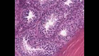
Histology of TESTIS

Вставка
- Опубліковано 15 жов 2020
- A capsule composed of dense collagenous material that encloses the testicle is called the tunica albuginea. Most species contain a vascular layer within the tunica albuginea. Horses often contain smooth muscle fibers within the tunica albuginea. The double layer of simple squamous epithelium and mesentery directly covering the testes and apposed to the tunica albuginea is the tunica vaginalis.
The parenchyma of the testicle is divided into lobules by loose connective tissue bands (septuli testes). These lobules are composed of tubules lined by stratified epithelium composed of maturing germ cells (spermatogonia, spermatocytes, spermatids and spermatozoa) and Sertoli cells. These tubules are supported by a basement membrane which contains fibroblasts and myofibroblasts. The Sertoli cells are triangular or oval in shape with a prominent nucleolus and fine chromatin. These tubules (seminiferous tubules) constitute the exocrine portion of the testes. The maturing germ cells are stratified so that the spermatogonia constitute the basilar compartments closest to the basement membrane. The spermatocytes, spermatids and spermatozoa constitute the next layer or the apical compartment with the spermatocytes adjacent to the basilar compartment and the spermatozoa closest to the lumen.
The endocrine portion of the testes is comprised by the Leydig or interstitial cells which are present between seminiferous tubules primarily located in the extra-tubular connective tissue. These cells are responsible for testosterone secretion.
As spermatozoa mature in the seminiferous tubules, they are guided out of the testes by the rete testis (randomly arranged) or tubuli recti (straight tubules) which are connected to the seminiferous tubules. These tubules eventually lead to the extragonadal tubular structures.


Exams next week and ur videos made my studies much simpler and easy to remember ❤❤
U made learning histology easy mam..tq mam
Thank you😊🙏
Nice video.
Thank you 😊
Well explained but a little bit more explanation is needed anyways it's nice 😀👍 I've shared with my students
I'm confused as to why is tunica albuginea the most outer layer? Every diagram I have seen says the most outer layer is tunica vaginalis. I'm not sure if this is a mistake or I misunderstood. Here I copied the last text: "Each testicle is covered by tough, fibrous layers of tissue called the tunica. The outer layer is called the tunica vaginalis and the inner layer is called the tunica albuginea."
As per I remember tunica vaginalis is a vascular layer and it is inner layer and albugenia Is always outer
Tunica vasuculosa is the vascular layer@@gamingstation4801
We can consider it like this, basically we assume testis has three layers (tunica vaginalis, albuginea, vasculosa) from out to in. But originally tunica vaginalis is not the true part of testis rather it is a derivative from peritoneum, moreover posterior side of testis is devoid of tunica vaginalis ( instead we have thickening forming mediastinum testis inside), so in histology we consider tunica vaginalis as outer layer. Hope it helps
Wahiyat explanation 🎉🎉
Thank you mam for this video🙏, waiting for more videos 😘😘
You're welcome 😊
drive.google.com/file/d/18eehoG_8cp2Tp8TDSvGATv2dyqkRMs3a/view?usp=drivesdk
@@Dr.KareemaTabassum thank u so much mam...I m pathology pg resident.these slides helps to revise the basic things
Thanks!
You're welcome 😊
ua-cam.com/video/_jkNWGLSNxA/v-deo.html
Nice video
Thank you 😊
Very good explanation, much appreciated. The music feels a little loud tho
Amazing explanation
Thank you 😊
Thank you
Very nice thanks mam
Thank you 😊❤
Thank you ma’am
Great explaination, please remove music
Amazing 👏
Thank you so much 😍😍
You're welcome 😊
🔥🔥🔥
Nice explaination👌
Thank you 😊
Can I learn like this in bsc 2nd year
👍👍😇😇
What cells surround the outside of seminiferous tubule in a circle?
Smooth muscles
❤
Okay so u have made my anat half easy 😄
That means alot❤️
Nice video
Thank you 😊