Thank you very much for this amazing ressource. No book or other video I can think of could have shown this in a more understandable and practical way! Much appreciation from Berlin
This was so helpful. As a nurse just starting in the cath lab this explained so much that people just assume you know. This makes me feel so much more confident. Thanks so much!
Excellent Video and the best way to learn the Angio views. I had a doubt - Like you mentioned - When the Diaphragm will be visible in a Cranial View. Similarly, what is the landmark to identify if the View is a Caudial View ?
Thank you for sharing! Very helpful! Do you have a good resource with good examples of LHC with each coronary artery identified? I'm starting my cardiology fellowship in July and I found your videos very helpful!
Deya Alkhatib. Thanks for watching the videos. I am glad you find these helpful. For diagnostic LHC, views, coronaries, techniques and complication Morton Kern is a good resource. Hers the link to the book www.amazon.com/gp/aw/d/0323597734/ref=dp_ob_neva_mobile
Hi Christina I just offered a position in Cath Lab… excited but super nervous. 5 years of ICU experience. Definitely feel like this will be a new learning curve for me. How is you experience is going in the Cath Lab
Great video! One suggestion i would make would be to include anatomical structures in the views to make it easier to identify like spinal, sternal and descending aorta location.
Hi thanks for the cours , i have a question please , i want to know if the image on the camputeur is the image on the intensifier ( point of view = image intensifier) or the point of view is the x ray source, in other terme if we look to the heart from the x ray source or we just look at the ember of the x rays on the intensifier thanks
Amar. The image on the computer is if you are looking at the heart from the X-ray source. Imagine if the X-ray source is your eye, heart is an object you are looking and the final image on the computer will be the shadow of that object on the wall (in this case on the image intensifier and computer). If you get this concept, the arteries as well as other mediastinal structures orientation will be start making sense. Hope this help..
i ask because, in the front view, we see the spine in the left side of the camputer, and the axis of the heart to the left, while if the our eyes = the x ray ( coming from bellow) , we should see the spine on the right side and the axis of the heart to the left ( I hope you understood me)
This is amazing. As a training cardiac radiographer, I now have an Idea on the importance of those views with corresponding anatomy. Can you also explain the plain ap cranial and caudal views for LCA? Thank you so much.
Thanks a lot sir, it's really wonderful explanation. Is there any particular degree regarding how much of LAO /RAO and Caudal/ cranial tilt to be taken while doing coronary angiography?
medikonda Parameshwara Reddy.!Thanks for the nice comment. Usually its best to start with an angle between 35-45 for LAO/RAO as well as for caudal/cranial. Depending on the first image, you can always adjust the angle depending on the patient body habitus and orientation of the heart. If you go shallow (low angles) the arteries will be overlapped. If you go very steep (More angle) you will be able to separate the arteries but at the cost of 1. More radiation to the patient. 2. More radiation to yourself give scattered beam. 3. Decrease image quality. I hope this help
Please guys i want help I cath lab technologist and I am new to the job and i just want a guide on how to provide the best image includin the panning of the table
very useful... thank you for that wonderful video... i wonder why few likes for this video.. EXCELLENT SIR,, what ddevice/software you use to make whiteboard drawings like this ???
Thanks for the nice comments. These lecturers are just to give students an overview on cardiology topics in a very basic and practical way and there is nothing more rewarding than having them understand these difficult topics. Feel free to share these with anybody. I use AZ screen recorder to record and autodesk sketchbook for presentation. I hope this help.
Suvendu Maji. Thanks for watching. LAO caudal view will be good to view the proximal/ostial segments of both Ramus and LAD. For the rest of the LAD you may choose some cranial view and for Ramus a caudal view. Hope this help.
Terrific didactic video!! However your comment box was obscuring the image when you were showing the pathologies. Kindly see if you can "circumvent" it! Regards, Dr Ashish Chakravarty
neetesh agrawal. I will cover it separately but for the body of RCA straight LAO or Caudal is good. RAO for shortens the body of RCA. For the distal bifurcation and branching vessels, AP cranial and RAO cranial views are good. Hope this help.
Lavisha Gandhi, thanks for your comments. Unfortunately, I dont have written notes for these. All these lectures are live and prepared from the information gathered from the memory. I am sorry.


Thank you so much for this I don't mean to repeat the comment before me but also a cath lab nurse, really appreciate this.
Such a brilliant helpful video. Thanks for posting
Thank you very much for this amazing ressource. No book or other video I can think of could have shown this in a more understandable and practical way! Much appreciation from Berlin
Thanks for the nice comment and thanks for watching. Glad these are helpful. Stay blessed
Thanks a ton. Very clearly explained. Simply wow...
Thank you for the great videos! Keep it up!!!
Excellent overview. Thank you!
Thank you so much.Very useful video.May God bless you sir❤
This was so helpful. As a nurse just starting in the cath lab this explained so much that people just assume you know. This makes me feel so much more confident. Thanks so much!
Thanks for the nice comment. Much appreciated
Thank you very much for your kind effort. I really helped me a lot.
Best basic angio tutorial I’ve came across
Appreciated. Thanks for watching
Thanks for mind blowing excellent presentation in CAG.
Thanks for watching and the nice comment. Appreciated
Beautiful talk.
Huge ! Thanks a lot - very helpful indeed!
Nikolaus Hauser. You are welcome. Thanks
Thanks for your videos. Very helpful. Keep it up.
Thanks for watching. Glad these are helping. Appreciated
Excellent Video and the best way to learn the Angio views.
I had a doubt - Like you mentioned - When the Diaphragm will be visible in a Cranial View. Similarly, what is the landmark to identify if the View is a Caudial View ?
Thank you very much for another great video
A Alsherbini thanks. You are welcome..
Superb presentation and teaching..VERY GOOD BROTHER...
Thank you sir 😇😇😇
Thanks for watching and the nice comments. Appreciated
1000% helpful! Thank you!!!
A. L. Thanks a lot. Glad these are helpful..
Thank you for sharing! Very helpful! Do you have a good resource with good examples of LHC with each coronary artery identified? I'm starting my cardiology fellowship in July and I found your videos very helpful!
Deya Alkhatib. Thanks for watching the videos. I am glad you find these helpful. For diagnostic LHC, views, coronaries, techniques and complication Morton Kern is a good resource. Hers the link to the book
www.amazon.com/gp/aw/d/0323597734/ref=dp_ob_neva_mobile
Hi Christina I just offered a position in Cath Lab… excited but super nervous. 5 years of ICU experience. Definitely feel like this will be a new learning curve for me. How is you experience is going in the Cath Lab
Great video! One suggestion i would make would be to include anatomical structures in the views to make it easier to identify like spinal, sternal and descending aorta location.
Thanks for watching. I think this is a great idea. Will try to add this in the future videos.
Excellent help for a new cath tech. too. Great video.
Thanks for watching and the nice comment
Hi plz can you share with me any data for cath tech like how to move the table to center the tip in every. View
@@alwoyibrahim5280 knowing anatomy and machinery is you best bet. No real tips other than practice panning. For LHC always put the tip at 11'oclock.
Nice n precise....Very effective.
Thanks Abdullah. Glad this is helpful..
Thank you very much for the video it is clear and concise :)
Thanks a lot and thanks for nice comments. I am glad these are helping…
Thank you so much! It was so helpful!
Thanks for watching. Glad it was helpful..
Excellent
Your hand writing is so beautiful, i wish i could have 😅
Very helpful. Thank you.
Thnx for watching…
thank you from Indonesia❤️
Godong Salam. Appreciated. Regards from USA 😀
Hi thanks for the cours , i have a question please , i want to know if the image on the camputeur is the image on the intensifier ( point of view = image intensifier) or the point of view is the x ray source, in other terme if we look to the heart from the x ray source or we just look at the ember of the x rays on the intensifier thanks
Amar. The image on the computer is if you are looking at the heart from the X-ray source. Imagine if the X-ray source is your eye, heart is an object you are looking and the final image on the computer will be the shadow of that object on the wall (in this case on the image intensifier and computer). If you get this concept, the arteries as well as other mediastinal structures orientation will be start making sense. Hope this help..
@@whiteboardandmarkercardiol2787 thank you so much, it will be very usefull
i ask because, in the front view, we see the spine in the left side of the camputer, and the axis of the heart to the left, while if the our eyes = the x ray ( coming from bellow) , we should see the spine on the right side and the axis of the heart to the left ( I hope you understood me)
This is amazing. As a training cardiac radiographer, I now have an Idea on the importance of those views with corresponding anatomy. Can you also explain the plain ap cranial and caudal views for LCA? Thank you so much.
Thanks a lot and thanks for watching. Will cover that too
Hi I also like you but what i want to know on how to provide best image the key setup of the tip of catheter and the panning of the table
Vert helpful... thank you
Thanks for watching..
Thanks a lot sir, it's really wonderful explanation. Is there any particular degree regarding how much of LAO /RAO and Caudal/ cranial tilt to be taken while doing coronary angiography?
medikonda Parameshwara Reddy.!Thanks for the nice comment. Usually its best to start with an angle between 35-45 for LAO/RAO as well as for caudal/cranial. Depending on the first image, you can always adjust the angle depending on the patient body habitus and orientation of the heart.
If you go shallow (low angles) the arteries will be overlapped. If you go very steep (More angle) you will be able to separate the arteries but at the cost of 1. More radiation to the patient. 2. More radiation to yourself give scattered beam. 3. Decrease image quality. I hope this help
@@whiteboardandmarkercardiol2787 thanks a lot for clarification. Can you make videos on Basic steps in PCI.
Thanks, very helpful
Appreciated. Thanks for watching
Thanks
Thank you!!
You are welcome. Thanks for watching...
Please guys i want help I cath lab technologist and I am new to the job and i just want a guide on how to provide the best image includin the panning of the table
Thank you so much
Thanks for watching
thank you!
Thanks for watching
Thankyou so much sir.....👍
Thnx for watching
Thank you sir
You are welcome.. thanks
very useful... thank you for that wonderful video... i wonder why few likes for this video.. EXCELLENT SIR,,
what ddevice/software you use to make whiteboard drawings like this ???
Thanks for the nice comments. These lecturers are just to give students an overview on cardiology topics in a very basic and practical way and there is nothing more rewarding than having them understand these difficult topics. Feel free to share these with anybody.
I use AZ screen recorder to record and autodesk sketchbook for presentation. I hope this help.
@@whiteboardandmarkercardiol2787 how to you draw on screen ?? surface pro ??
@@whiteboardandmarkercardiol2787 i just shared your videos in the indian cardio pgs group.
I use a Samsung Android Tablet.
Thanks for sharing it. Always happy to help and encourage students to learn more about cardiology.
I have added more videos on coronary angiogram, views, anomalies and quantification. Feel free to share with anybody/forum/pages who can benefit....
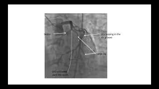
Sir If i want to focus on LAD /Ramous intermediate which one view will be better?
Suvendu Maji. Thanks for watching. LAO caudal view will be good to view the proximal/ostial segments of both Ramus and LAD. For the rest of the LAD you may choose some cranial view and for Ramus a caudal view. Hope this help.
Very helpful
Thanks. Appreciated
Thank u sir
deepak kumar Rai. You are welcome
Terrific didactic video!! However your comment box was obscuring the image when you were showing the pathologies. Kindly see if you can "circumvent" it! Regards, Dr Ashish Chakravarty
Thanks for the nice comment and thanks for watching. Will make sure to edit videos in future. Thanks
Sir for RCA which view is better
neetesh agrawal. I will cover it separately but for the body of RCA straight LAO or Caudal is good. RAO for shortens the body of RCA.
For the distal bifurcation and branching vessels, AP cranial and RAO cranial views are good.
Hope this help.
I want written notes of coronary views u have explained very well it's very useful but if u provide wriiten notes
It's also better
Lavisha Gandhi, thanks for your comments. Unfortunately, I dont have written notes for these. All these lectures are live and prepared from the information gathered from the memory. I am sorry.
Okay sir no problem thanks a lot for sharing this live lecture ❤️🙏❤️
Sir is there any way to communicate with u
Sure. You can chat here publicly or privately. Good luck
@@whiteboardandmarkercardiol2787 sir can I get a chance to study CATHLAB on US.
Right heart catheterization
Here you go.. thanks for watching
ua-cam.com/video/ZLJXCxq3Au4/v-deo.html
Spider view is not drawn correctly
Thank u sir
You are welcome. Thanks for watching...