Structure of human Eye: Human Eye Anatomy

Вставка
- Опубліковано 11 гру 2024
- Welcome to My UA-cam Channel Power of knowledge Academy. In This video you will learn about the detailed structure of human Eye or Anatomy of Human Eye.
The Human Eye
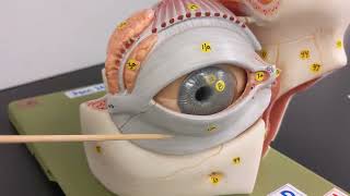
The human eye is a complex and highly specialized organ responsible for vision. It is capable of perceiving light and converting it into electrochemical impulses that the brain interprets as images. The anatomy of the eye can be broadly divided into external structures, optical components, and internal structures.
External Structures:
1. Sclera: The white part of the eye, a tough, fibrous layer that protects the inner components and provides attachment points for the muscles that move the eye.
2. Cornea: A transparent, dome-shaped surface at the front of the eye that helps to focus incoming light onto the retina.
3. Conjunctiva: A thin, transparent membrane that covers the sclera and the inner surfaces of the eyelids, providing a protective barrier.
4. Eyelids and Eyelashes: Protect the eye from debris, dust, and excessive light. Eyelids also help spread the tear film over the eye, keeping it moist.
5. Lacrimal Apparatus: Includes the lacrimal glands (which produce tears), lacrimal ducts (which distribute tears over the surface of the eye), and nasolacrimal ducts (which drain tears into the nasal cavity). This system keeps the eye lubricated and helps remove foreign particles.
Optical Components:
1. Iris: The colored part of the eye, which contains a circular aperture (pupil) in the center. The iris regulates the amount of light entering the eye by adjusting the size of the pupil.
2. Lens: A transparent, flexible structure located behind the iris, capable of changing shape to focus light onto the retina.
3. Aqueous Humor: A clear fluid that fills the space between the cornea and the lens, providing nutrients and maintaining intraocular pressure.
4. Vitreous Humor: A gel-like substance that fills the space between the lens and the retina, helping to maintain the eye's shape and optical properties.
Internal Structures:
1. Retina: A thin layer of tissue lining the back of the eye, containing photoreceptor cells (rods and cones) that convert light into neural signals.
2. Optic Nerve: Transmits visual information from the retina to the brain.
3. Macula: A small central area of the retina responsible for high-acuity vision, where the concentration of cones is highest.
4. Choroid: A layer of blood vessels between the retina and the sclera, providing oxygen and nutrients to the eye.
5. Ciliary body: The ciliary body is a ring-shaped structure in the eye that produces aqueous humor and contains muscles which adjust the lens for focusing.
6. Optic Disk: The point on the retina where the optic nerve fibers exit the eye, known as the blind spot because it lacks photoreceptors and is insensitive to light.
To know detail about each structure of human eye, watch the complete video and share your comments on topic.
#HumanEyeAnatomy
#VisionScience
#OcularBiology
#RetinalStructure
#OpticNerveFunction
#IrisAndPupil
#CornealLayers
#LensAndFocusing
#VisualAcuity
#EyeHealth


![LAVROV's interview with Tucker CARLSON 😁 [Parody]](/img/n.gif)
Well explained sir
Sir you explain great....!!!!
Really I'm wait for this lecture thank you sir 💙
Sir ap k lectures or diagrams 2no hi bohat outstanding hoti hh ❤
Shukriya bhot hi accha samjhaya aapne.
Notes bhi share krte toh accha hotaa...❣️🇮🇳
Very helpful video
Bhot acha vedos hy sir
Nice explanation
Zabardast tareeeen 🎉
❤❤❤❤❤❤❤❤❤❤
❤❤❤❤❤❤❤❤❤❤
Method of teaching is nice
Best explained sir, I saw many videos on this topic all were good but yours is most well explained thanks
Super sirji
Sir your drawing your explanation superb
Really good video quality
Your drawing is so beautiful
Minimum time me achha knowledge diya aapne thans brother
Love from India 🇮🇳
Thank you sir 😊
wow welldone sir i appriecate you you become a good or excellent professor...😊❤
Osam
Thanks 😊
Excilent sir pr aap sabhi part ko ट्रांसलेसंस karke padhna sir
Hello sir.. Could you plz explain about glaucoma of eye????
Bohot helpful
Excellent
Best vedio
Brilliant
Well explained sir ❤
Superb sir🤗🤗 please video k end me short notes v provide kr dijiye sir please 🙂
Video bahut acha hai..... Digital bord
Well done 👍
❤❤❤
Sir please an lecture on the dialysis
When will you upload Next video
Thanks sir
Perfect lacture
Can you give concept about cillary muscle
Yooo sir is op❤
Sir please make a video on DNA fingerprinting.....
Nice video for explaining eye anatomy but muje ek chij clear ni hui, first diagram me Iris and pupil ke bich me jo white part h use kya kehte hain??? Pupil se just upar aur iris k andar...agar kisi ko pata h then reply....
😊😊😊
Sir ligation py aik lecture Dy dain plzz
Sir plz upload lectures biology class 12 for new session from new book
Mashaallah ❤
Sir G vitamins ke licture upload kre
Assalam o alaikum sir
Sir kia ap malprsentation and malprsentation clear krwa dien gay
Your video is very good but please speak in English. You’ll get more views worldwide
😢agey hi hum shukr krte hain kay Urdu me itne ache sir mile hain English me bus paper dena chahei baqi lecture Urdu me good hai zra angrezo ko bhi pta chle
Sir, please teach English 🙏
Sir This we study in class 8 and 10 in Indian
Assalamualaikum sir aap bsc ki class chalu kar sakte hai kya please Bsc zoology and botany ki class.
Jai shri ram
Masaallha ❤❤❤
Bhot acha vedos hy sir