Fantastic primers for histology, as a med student I really appreciate you taking the time to make these and share them with the public! Thanks so much!
The original reason that the epithelium lining the lower urinary tract was called "transitional" is that it "changes" in thickness during, say, the contraction and relaxation of the bladder. Today, it is more correctly called "urothelium"
One year ago i studied this video and got a similar sample at my exam, I succeeded thanks to you but had never come back to say thank you man ! Hope everything s good for you
"I'm trying not to use a lot of ESOTERIC words which really don't matter and which will confuse you..." I had to look up "esoteric" to find out what it meant.
@@SFGrim-hc3ci Many things evolve in medicine. But. Anatomy and pathology can’t simply be changed. Yes, maybe classifications and new things could be found. But those basics, they stay. I’m happy that those videos aren’t deleted for so much time.
So ur at 6th year of medicine , sorry to take a little of you're time but i want to ask u , I am in my 1th year of medicine and i have an exam in histology if u could tell me how do u stady histo for an exam??
you, Sir, are a wonderful human being and are certainly enriching the field of medicine with your knowledge and kindness! i have a histology exam coming up tomorrow and could not be more prepared
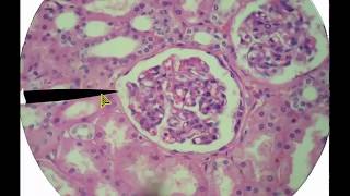
The part of the distal convoluted tubule in proximity with the vascular pole is the macula densa, which forms only part of the juxtaglomerular apparatus, no?
I really enjoy watching your videos to get an averview and to prepare for exams, they are great!!! Unfortunately this video is out of sync, can you fix that ?
@Punkomatul Oh? I thought the mesangial cells were sort of the connective tissue of the corpuscle, and that the extraglumerular ones were located in the vascular pole.
@Bahamut2218 intraglomerular mesangial cells are sort of a connective type of cell (though note that they're "sort of") from within the glomerule and extraglomerular mesangial cells are the ones at the vascular pole that we talked about earlier....i hope you understand what i mean :)
First off I'd like to say your vids are of great help to me. I would just like to remark that the (macula densa?) cells involved in the renin system you refer to at 4:30, are part of the distal (not the proximal) tubule....or I just didn't get it right :-)
I think there is a mistake at 4:32 when you mention that part of the Proximal convoluted tubule forms the macula densa. It should be the distal convoluted tubule.
Thank you for this video. Are the cells that lines the capillaries called ENDOTHELIAL cells rather than epithelial cells. Epithelial cells are usually for protection. Endothelial cells are specialized for the exchange of nutrients and oxygens, and toxins, from the blood. I need this verified. Thanks. Brilliant Lecture.
The easiest way o tell the DCT(Distal convoluted tube) lacks the brush border so the inside of the tubule will have next to nothing in the center, while the proximal has the brush border so in the center there will apear cloudy or occluded. Hope that helps.


Fantastic primers for histology, as a med student I really appreciate you taking the time to make these and share them with the public! Thanks so much!
The original reason that the epithelium lining the lower urinary tract was called "transitional" is that it "changes" in thickness during, say, the contraction and relaxation of the bladder. Today, it is more correctly called "urothelium"
One year ago i studied this video and got a similar sample at my exam, I succeeded thanks to you but had never come back to say thank you man ! Hope everything s good for you
"I'm trying not to use a lot of ESOTERIC words which really don't matter and which will confuse you..."
I had to look up "esoteric" to find out what it meant.
In my 6th year of medicine and these videos help a lot! Thank you for your amazing work!
that fact that these video's being as viable as they are at almost 20 years old now is crazy.
@@SFGrim-hc3ci Many things evolve in medicine. But. Anatomy and pathology can’t simply be changed. Yes, maybe classifications and new things could be found. But those basics, they stay. I’m happy that those videos aren’t deleted for so much time.
So ur at 6th year of medicine , sorry to take a little of you're time but i want to ask u , I am in my 1th year of medicine and i have an exam in histology if u could tell me how do u stady histo for an exam??
you, Sir, are a wonderful human being and are certainly enriching the field of medicine with your knowledge and kindness! i have a histology exam coming up tomorrow and could not be more prepared
yup, u right, i screwed up, i OFTER screw up, thanks for being a keen listener
Thank you so much sir...The video is really so much helpful
proximal so packed with microvilli, it looks like it has no lumen
Please fix the audio. Thank you so much dr. Minarcik.
The audio is out of sync, seems to be ahead of video
At the beginning the slide looks very happy.
"epithelium" is a generic term used for any flat lining cell such as mesothelium or even synovium
I am impressed. You are such a great help. May God bless you with a long heathy life
Thank you very much for sharing this video, you are making things soo much easier for histology
Thank you, your videos were helpful and I passed my exam!
Hehe, rumors of my death have been greatly exaggerated!
These videos are all unbelievably helpful
The part of the distal convoluted tubule in proximity with the vascular pole is the macula densa, which forms only part of the juxtaglomerular apparatus, no?
@WashingtonDeceit; Harbinger631 is right the audio is out of sync. If you have time can you please fix it? This video is very valuable - thank you!
I really enjoy watching your videos to get an averview and to prepare for exams, they are great!!!
Unfortunately this video is out of sync, can you fix that ?
Excuse me professor, why didn't you talk about the medulla?
Thank YOU! Now i understand Histology!!! U save my live!!!
@Punkomatul Oh? I thought the mesangial cells were sort of the connective tissue of the corpuscle, and that the extraglumerular ones were located in the vascular pole.
@Punkomatul And those are called juxtaglomerular cells, from what I've learned.
Mesangial cells are visceral, He says that towards the beginning but then calls them parietal near the end.
great work you help a lot with my slide test final.but why don't you mention the loop of henle in the medulla?
@Bahamut2218 yes indeed, the other part is composed of smooth muscle fibres from the arterial walls that have suffered secretory differentiation.
@Bahamut2218 intraglomerular mesangial cells are sort of a connective type of cell (though note that they're "sort of") from within the glomerule and extraglomerular mesangial cells are the ones at the vascular pole that we talked about earlier....i hope you understand what i mean :)
Really good that u r making these movies!
BUT on this movie the voice does not synchronize with the movie... :)
First off I'd like to say your vids are of great help to me. I would just like to remark that the (macula densa?) cells involved in the renin system you refer to at 4:30, are part of the distal (not the proximal) tubule....or I just didn't get it right :-)
I think there is a mistake at 4:32 when you mention that part of the Proximal convoluted tubule forms the macula densa. It should be the distal convoluted tubule.
@Bahamut2218 yeah i belive that's correct. You can also call them extraglomerular mesangial cells if you want.
shotgun histology, you champion
the audio is out of sncy for most of the second half of the video
Such amazing video. Thanks sir!
I dont know if its just my comp,..but it seems that the timing is off..
The sound is a little bit off, right?
Mazing video - But can someone tell me how to spot and tell the difference between proximal/distal tubules?
You sir are a legend
Moath Abdulghani Still Alive too!
WashingtonDeceit legends never die :)
Moath Abdulghani You made my day!
Thank you for this video. Are the cells that lines the capillaries called ENDOTHELIAL cells rather than epithelial cells. Epithelial cells are usually for protection. Endothelial cells are specialized for the exchange of nutrients and oxygens, and toxins, from the blood. I need this verified. Thanks. Brilliant Lecture.
I love this guy.
Its worth watching this video but still i don't get how to differentiate proximal and distal tubules. Both looks similar.
The easiest way o tell the DCT(Distal convoluted tube) lacks the brush border so the inside of the tubule will have next to nothing in the center, while the proximal has the brush border so in the center there will apear cloudy or occluded.
Hope that helps.
So nice and simple...thanks a ton!!!
i agree with you as there is mistake on the end of
perhaps you can listen to some mccain speeches?
edit the video sound is 20 sec earlier than video!!
this video should definetely be re-recorded...
Thank you so much this video helped me alot
Lifesaving! thank you so much!
Love this video as well thanks for sharing with us ;D Blessings
Thank you
thank u sir. u really helping
At scale of one to ten you are awesome 17!
:)
this is so confusing. :( im lost. its not synchronized i dont think. because i dont see anything youre talking about.
This is so helpful, thank you!
MY SAVIOUR!
thank you sir
sound isn't synched with the video... unfortunately :(
Yes, you are correct. :)
university of iowa's virtual histology website
Painless kidney
Hi all RSU international students reading for tomorrows Histology Colloquium
voice is faster than the mouse.. i'm getting confused
very very very good
holla to all kcl students with the spot test tomorrow
2:23-3:04
Sen adamsın türkler likelsasın
thanx but voice out of sync
The sound isn't synched with the video, which defeats the purpose of the video. If that could be fixed this video would be amazing.
I love you, you're the best, can I have your babies?
stop smoking weeed!!! ur mixing too many stuff :D
you must be blind then
If you need some help before an exam just google histology.be and you will find an electronical microscope.
A big thanks from belgium
mr your voice is so so so so so looooooow please fix it.thank you.