Це відео не доступне.
Перепрошуємо.
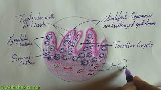
Histology of Palatine tonsil \Tonsil

Вставка
- Опубліковано 26 жов 2020
- Tonsils
Tonsils belong to the mucosa associated lymphoid tissue (MALT), but because they are incompletely encapsulated, they are considered organs and will be studied apart from the MALT. The tonsils constitute a lymphoid tissue that lies beneath, and in contact with, the epithelium of the initial portion of the digestive tract. Depending on their location, tonsils in the mouth and pharynx are called palatine, pharyngeal, or lingual.
Palatine Tonsils
The two palatine tonsils are located in the lateral walls of the oral part of the pharynx. They are lined with a squamous stratified epithelium that often becomes so densely infiltrated by lymphocytes that it may be difficult to recognize. The lymphoid tissue in these tonsils forms a band that contains free lymphocytes and lymphoid nodules, generally with germinal centers. Each tonsil has 10-20 epithelial invaginations that penetrate the tonsil deeply, forming crypts, whose lumens contain desquamated epithelial cells, live and dead lymphocytes, and bacteria. Crypts may appear as purulent spots in tonsillitis. Separating the lymphoid tissue from subjacent structures is a band of dense connective tissue, the capsule of the tonsil. This capsule usually acts as a barrier against spreading tonsillar infections
the numerous nodules that compromise the palatine tonsil.
Lymph Nodules - spherical aggregations of lymphocytes that usually have germinal centers.
Crypts - infoldings of the epithelium into the underlying connective tissue.
Lymphocytes pass through the epithelium in areas of inflammation.
Lymphocytes are seen in the lumen of some crypts.
Sequestered crypts are usually inflamed and filled with debris and lymphocytes (pus).
Plasma Cells - large numbers of plasma cells are usually seen in the underlying connective tissue near the epithelium.


Ur Videos are Boon ✨ for me in such a stressful situations ... excellent coverage in less time
Thank you, means alot 😊
This is just what I needed. Good explanation!
Very clear explanation
Thank you ma'am, it would really help me for my presentation on this topic.
Ur just doing amazing ..... Really very helpful thank you thank you so much these videos really helped me during my practical exams all bcoz of u 💕💕♥️♥️♥️
Thank you .........
You're welcome 😊
Love you mam awsm ur saviour U voice is also totally melody awsm quality content
I will like your each and every video blessed to see ur video
Thanks a lot 😊
Very nice explanation
Thank you...😊
Thank you mam... awesome explanation mam
You're welcome 😊
thank you for your explanation:) may i know the references teory about histology palatine tonsils :) ? thank you ;)
You're welcome 😊, I took reference of diagram from online and theory from IB Singh
Thank u mam it was usefull🤗
Clear
Explanation 🔥🔥
Thank you,❤️
I need Lymphocyte labelled diagram...do you have this? Can you please upload or send me.. Please?
Tomorrow is my university exam and I am watching now😅😅
Same bro!!!
How did your exam go?
All the best🌹
@@Dr.KareemaTabassum very well
Same
Plz make videos on remaining topics of anatomy 😢
Plz ye music na lagaya kren peeche
Please give subtitles and don’t use music .