Advanced Ultrasound Cervical Anatomy

Вставка
- Опубліковано 26 вер 2024
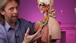
- We demonstrate how to establish ultrasound views for the supraclavicular and interscalene approach to the brachial plexus. We also demonstrate tracing of individual nerves roots to their respective neuroforamen for accurate identification of brachial plexus components and performance of procedures including selective extraforaminal cervical root blocks and stellate ganglion blocks. Skeletal model images supplied courtesy of Complete Anatomy from Elsevier.
Additional reading:
Martinoli C, Bianchi S, Santacroce E, et al. Brachial plexus sonography: a technique for assessing the root level. AJR Am J Roentgenol. 2002;179(3):699-702
Procedure performed by Raf Vazquez, MD (@RafMD1) and David Hao, MD (@davidhaomd). Peer reviewed by Charlie Kelly, MD.
--
Note that the orientation of the ultrasound image may vary by institution and resource.
Equipment:
GE Logiq E Ultrasound Machine
--
Invasive medical procedures can result in harm to patients and practitioners and should be performed only by qualified medical professionals. This video is intended solely for informational purposes and to supplement, not replace, proper training and supervision by qualified instructors.
Viewers are advised to check the most current information provided by the manufacturer for every device being used, and to verify the indications, contraindications and proper procedural technique. The dose, method of administration, and contraindications for any administered drug should be confirmed before use.
The authors of the video do not assume any liability for any injury or damage to any person or property arising from the use of this video.


Thank you 👍👍
In the first supraclavicular view: I think serratus anterior should be middle scalene muscle
Excellent
Is this competitive with MRI? At least it can be done upright.