Does the splenic vein really run along the inferior margin of the pancreas? I thought it ran along the superior margin, where it left a groove, and then move on the posterior face of the organ as it progressed towards the splenomesenteric trunk or portal vein
+Sana Lab one silly basic question and I'm even embarrassed to ask it but I just couldn't find ANY page to clarify when we look at structures on Ultrasound we see them from above don't we? if not then from are we looking is the saids? as in pregnancy ... or behind .. thanks in advance
Whatever you've typed and attempted to ask is extremely confusing. I'll just tell you what I know about the view from the screen to the body: The top of the image on the screen is the anterior-most portion. Moving down the screen towards the bottom, you're moving from the front to back of the patient (unless it's a trans-vaginal scan, that's a whole different story).
Hi, your question was not "extremely confusing" despite what someone commented! As the question was so long ago I'll assume you understand the concept but if not, ask.
Mild nonspecific gallbladder wall thickening in the absence of gallstones. Findings can be seen in the setting of hepatocellular dysfunction and right heart failure among multiple other etiologies. Additional workup as clinically warranted
Yes splenic vein exist the spleen and travels along the posterior border of the pancreatic tail and body while splenic artery enters the spleen at the splenic hilum superior and anterior to the splenic vein

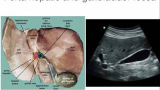


Thank you for reminding me how the Glisson's capsule and portal vein looks like under the echo view!
Best abdominal ultrasound explained video. I wanted to see the Liver because I have fatty liver. Thank you
Excellent lecture.
thank you for reminding me how the Glisson's capsule and portal vein looks like under the echo view!
Very good. Thanks.
Very Excellent and guiding lecture!
phenomenal, thanks
Excellent clarity. You are very knowledgeable.
Nice source of getting knowledge
Does the splenic vein really run along the inferior margin of the pancreas? I thought it ran along the superior margin, where it left a groove, and then move on the posterior face of the organ as it progressed towards the splenomesenteric trunk or portal vein
Thank so much for sharing Dr
Very helpful indeed really
Good video! :) thank you for your time.
Linda Carter Thank you for time taking for this view.
Very good information.
Nice and informative presentation
thank you
Thank you Doctor Beatrice Madrazo
+Sana Lab
one silly basic question
and I'm even embarrassed to ask it but I just couldn't find ANY page to clarify
when we look at structures on Ultrasound we see them from above don't we?
if not then from are we looking is the saids? as in pregnancy ... or behind ..
thanks in advance
Whatever you've typed and attempted to ask is extremely confusing. I'll just tell you what I know about the view from the screen to the body: The top of the image on the screen is the anterior-most portion. Moving down the screen towards the bottom, you're moving from the front to back of the patient (unless it's a trans-vaginal scan, that's a whole different story).
Hi, your question was not "extremely confusing" despite what someone commented! As the question was so long ago I'll assume you understand the concept but if not, ask.
Mild nonspecific gallbladder wall thickening in the absence of gallstones. Findings can be seen in the setting of hepatocellular dysfunction and right heart failure among multiple other etiologies. Additional workup as clinically warranted
Is it possible to see tumors that are in the gastrointestinal tract, if they’re big enough on an abdominal ultrasound?
Congratulation for compart your knowlege
tkank !
Thanks
👍🏻nice work
GOOD
Nice
INFORMATIVE
Y ARE YOU YELLING
I am having pain around my navel area. My ultrasound came back normal
Yes splenic vein exist the spleen and travels along the posterior border of the pancreatic tail and body while splenic artery enters the spleen at the splenic hilum superior and anterior to the splenic vein
Okay
Price
Hm
!!
1ㅣㅣㅣㅣㅣ11ㅣㅣㅣ1ㅣㅣㅣㅣㅣㅣㅣㅣㅣㅣㅣㅣㅣㅣㅣㅣㅣㅣㅣㅣㅣㅣㅣㅣ1111111ㄱ1
Ya brown boy😂😂😂
Thank you for reminding me how the Glisson's capsule and portal vein looks like under the echo view!
Thank you for reminding me how the Glisson's capsule and portal vein looks like under the echo view!
Thank you for reminding me how the Glisson's capsule and portal vein looks like under the echo view!