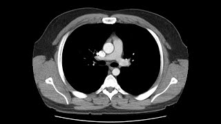
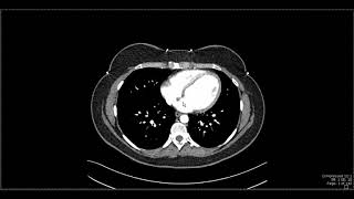
CT Cross Sectional Anatomy Abdomen (Axial View)

Вставка
- Опубліковано 29 чер 2024
- ➡️ LEARN MORE: This video lesson was taken from our Cross-Sectional Anatomy and Pathology course. Use this link to view course details and additional lessons. cloverlearning.com/courses/ct...
➡️ FREE TRIAL: Go to cloverlearning.com today to receive free access to hundreds of videos, quizzes, interactive learning tools, and our powerful certification exam prep tool!
➡️ JOIN OUR COMMUNITY:
/ cloverlearning
/ cloverlearning.inc
#cloverlearnining #radiology


what a piece of gold this video is!
thank you so much for this helpful video
Nicely explained!
Great video - many thanks!
Best CT abdomen concise lecture I have ever watched ;)
ua-cam.com/video/DqSG54aoufo/v-deo.html
what a great video, thanks. It really helps
That was a super helpful video!!! Thank you so much for posting it!!
ua-cam.com/video/DqSG54aoufo/v-deo.html
Great explanation
fantastic explaination...loved it totally
Thank you! This is so well explained
you can view more at ct.cloverleaflearning.com
Thank you very much, please do the same for the rest of the body, also perhaps MRI, and PET CT.
Thank you, and all the best.
ua-cam.com/video/DqSG54aoufo/v-deo.html
Well done dear doctor it was really very simple and so informative and great
you can view more at ct.cloverleaflearning.com
i have test soon thank u for this
WELL, EXPLAINED! 😃 I WAS CONFUSED WHEN I SAW THESE AS A ULTRASOUND STUDENT
Nicely explained
Thank you very much
Amazing...Thanks
Thank u so much
just wowww explaination
I have swollen inguinal lymph nodes. I can feel them but the ct scan says they aren't swollen. Why is that? Is it because the ct scan can't see it if it is smaller than 2 cm?
Thanks
Thank u so much Doc 🙏💕... Best 👍💯
You can get all 16+ hours of videos and more at www.cttechbootcamp.com
The descending aorta should be named as abdominal aorta below the vertebral level of T12.
Cn u add chest thorax anatomy...!
Plz make vedio with ideal procedure step by step
I have one question is the scanning mirrored.
So has the contrast not yet reached the v.cava?
very good!
Thanks. You can view more at www.radtechbootcamp.com
Colon or large intestine ulcers can be seen in ct scan?
Hi can you also make a chest and skull video.
Liver Spinal column aorta
Are we sure those were splenic veins? Cause they were enhanced when the descending aorta was and the IVC was not. Hmmmmmmm
Late arterial phase or portal phase between 40 seconds to 70 seconds after the Injection
What is the horizontal part in the middle between SMV,SMA and IVC , aorta (at 6:34)?
Left renal vein
3rd part of duodenum
You should be a teacher for teachers. Teach how to think and talk
🌼🌼🍀🍀❤