Ultrasound-Guided Arterial Line Insertion // Out-of-Plane Bevel-Guided Technique

Вставка
- Опубліковано 25 лют 2021
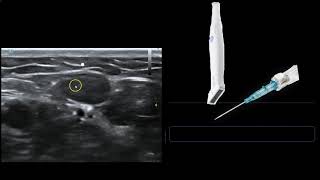
- We demonstrate how to perform an ultrasound-guided arterial line insertion with a 20 G IV catheter and the out-of-plane bevel-guided technique. This technique allows for continuous visualization of the needle from skin to vessel. We also demonstrate exchange of the 20 G IV catheter for an indwelling catheter via the Seldinger technique.
--
Technique demonstrated by Omar Hyder, MD.
--
Equipment:
Arrow Arterial Catheterization Kit
Custom Ref ASK-04125-MGH
GE Logiq E Ultrasound Machine
--
Disclaimer:
Invasive medical procedures can result in harm to patients and practitioners and should be performed only by qualified medical professionals. This video is intended solely for informational purposes and to supplement, not replace, proper training and supervision by qualified instructors.
Viewers are advised to check the most current information provided by the manufacturer for every device being used, and to verify the indications, contraindications and proper procedural technique. The dose, method of administration, and contraindications for any administered drug should be confirmed before use.
The authors of the video do not assume any liability for any injury or damage to any person or property arising from the use of this video.


That explanation of the angle of the transducer being perpendicular to the bevel is a game changer! Looking forward to trying it on my next a-line
Straight up
The ability to differentiate the shaft, bevel and the tip of the needle from each other (as explained at time point 5:00 in this vid) has made the OOP approach much, much safer and easier for me. Thanks for sharing that really awesome tip!
Truly this is the best U/S art line video on the internet, many thanks
Great vid! This is very practical and helpful than making many failed attempts and punctures
A RIJ tutorial is a must! Great video!
I like the idea of exchanging for a longer catheter - these always work better and don't damp out over longer term use vs shorter catheters.
Such fantastic content and available for free? Thank-you so much for this. The initial needle entry angle, along with the desired extent of dorsiflexion on the arm board, along with the decrease in attack angle on advancement of the needle will all reduce underlying wall puncture, however I appreciate covering said variables would prolong the video and perhaps warrant separate coverage. Thank you for making me a better clinician.
So clearly explained, well done
Thank you so much very good demonstration
Great explanation.
Too good explanation 👍
Great content!! Thank you
After you tilt the transducer to make the beam perpendicular to bevel, don't you lift it off the skin, losing the image?
You can maintain contact with the skin by applying pressure and ensuring sufficient gel. But yes, at a more extreme degree of angulation, you might lose the image.
@@PracticalAnesthesiaTechniques got it. Forgot to thank you so much for the great video, animation and response :) I had 2 more questions if you had time.
1) how far from the probe/what angle do you come at with the needle? Ie do you enter right at the transducer arrow or 1 mm in front if it?
2) when you're creeping forward with the probe, you talked about adjusting the needle left or right to center the probe above the vessel. Do you turn the probe as well or do you keep the probe the same way to keep the vessel centered? For example if you need to correct, the needle might be going diagonal to the probe instead of perpendicular.
Thanks again!!
Placing it like an IV is slick but I would always prefer to go 'through-and-through' for a higher success rate.
Success rate is a function of practice. If you keep practicing to place it like an IV and treat the artery like a vein (for which we would never go through and through), and use an appropriately shallow trajectory, you will find that you might fail a few times (like the medical student putting in their first few IVs), but you will eventually achieve 99.9% success. It's what you choose to embrace. However making 2 holes in a vessel if you don't have to doesn't make sense. May increase risk of long-term complications.