Animated Gross anatomy of Appendix: Position, Blood supply, Venous drainage, Nerve supply, Histology

Вставка
- Опубліковано 25 вер 2017
- 📌 𝐅𝐨𝐥𝐥𝐨𝐰 𝐨𝐧 𝐈𝐧𝐬𝐭𝐚𝐠𝐫𝐚𝐦:- / drgbhanuprakash
📌𝗝𝗼𝗶𝗻 𝗢𝘂𝗿 𝗧𝗲𝗹𝗲𝗴𝗿𝗮𝗺 𝗖𝗵𝗮𝗻𝗻𝗲𝗹 𝗛𝗲𝗿𝗲:- t.me/bhanuprakashdr
📌𝗦𝘂𝗯𝘀𝗰𝗿𝗶𝗯𝗲 𝗧𝗼 𝗠𝘆 𝗠𝗮𝗶𝗹𝗶𝗻𝗴 𝗟𝗶𝘀𝘁:- linktr.ee/DrGBhanuprakash
Animated Gross anatomy of Appendix - Position, Blood supply, Venous drainage, Nerve supply and Histology
The cecum and appendix are part of the gastrointestinal tract. They are the most proximal part of the large intestine, located between the ileum (distal small bowel) and the ascending colon.
Having served as a site for cellulose digestion in our ancestors, the cecum now simply acts as a reservoir for chyme which it receives from the ileum. The appendix contains a large amount of lymphoid tissue, but has no vital functions in the human.
In this article, we shall look at the anatomy of the cecum and appendix - their anatomical structure and relations, neurovascular supply and lymphatic drainage.
Anatomical Structure and Relations
The cecum. Note the blind end inferiorly, and its continuity with the ascending colon superiorly.
The cecum. Note the blind end inferiorly, and its continuity with the ascending colon superiorly.
The cecum is the most proximal part of the large intestine and can be found in the right iliac fossa and suprapubic region. It lies slightly inferior to the iliocaecal junction, and can be palpated in the right iliac fossa if enlarged due to faeces, gas or malignancy.
The name is derived from the Latin word ‘caecus’, meaning ‘blind’, due to its blind-end inferiorly. Superiorly, however, the open end of the cecum is continuous with the ascending colon. Unlike the ascending colon above it, the cecum is intraperitoneal.
The appendix, also known as the vermiform (worm-shaped) appendix is a narrow, blind ended tube attached to the posteromedial end of the cecum. The position of the free-end of the appendix is highly variable, and can be categorised into seven main locations. The most common positions are retrocaecal and subileal. A simple way to remember the positions is by imagining the appendix as the hour hand of a clock:
Pre-ileal - Anterior to the terminal ileum - 1 o’clock.
Post-ileal - Posterior to the terminal ileum - 2 o’clock.
Sub-ileal - Parallel with the terminal ileum - 3 o’clock.
Pelvic - Descending over the pelvic brim - 5 o’clock.
Sub-caecal - Below the cecum - 6 o’clock.
Paracaecal - Alongside the lateral border of the cecum - 10 o’clock.
Retrocaecal - Behind the cecum - 11 o’clock.
Neurovascular Supply
-----------------------------------
The cecum is derived from the embryologic midgut - and therefore blood is supplied by a branch of the superior mesenteric artery. The branch is the ileocolic artery, which splits into anterior and posterior caecal arteries to supply the cecum. Venous drainage is provided by the ileocolic vein, which empties into the superior mesenteric vein.
The appendix receives its blood supply via the appendicular artery (derived from the ileocolic artery), and drains through the appendicular vein. Both are contained, along with lymphatic vessels and nerves, within the mesoappendix, a fold of mesentery which suspends the appendix from the terminal ileum.
The autonomic nervous system innervates the cecum and appendix. It achieves this by means of the ileocolic branch of the superior mesenteric plexus, which follows the same course as the ileocolic artery.
The cecum, appendix and ascending colon. Note the inferior position of the cecum in relation to the ileum.
Arterial supply to the cecum and appendix, via the ileocecal artery.
Lymphatic Drainage
-------------------------------
Lymph from the cecum and appendix ultimately drains into the upper and lower ileocolic lymph nodes, which surround the ileocolic artery.
However, lymph from the cecum travels via a number of intermediate mesenteric nodes, whereas that of the appendix travels via a single intermediate node in the mesoappendix.
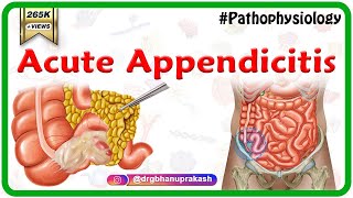
Clinical Relevance: Appendicitis
---------------------------------------------------
Appendicitis is acute inflammation of the appendix, and is the most common cause for acute, severe abdominal pain. The abdomen is most tender at McBurney’s point - one third of the distance from the right anterior superior iliac spine to the umbilicus. This corresponds to the location of the base of the appendix.
The aetiology depends on age. In the young, it is mostly due to an increase in lymphoid tissue size, which occludes the lumen. From 30 years old onwards, it is more likely to be blocked due to faecal matter.
Initially, the appendix cannot drain, and so increases in size, stretching the visceral peritoneum. This causes a vague pain in the periumbilical region. As the appendix swells, it irritates the parietal peritoneum, and causes severe pain in the right lower quadrant.
If the appendix is not removed, it can become necrotic and rupture, resulting in peritonitis (inflammation of the peritoneum).


Medvizz Animated medical video lectures :
1200+ complete animated medical Video lectures includes all high-yield topics enough to cover all contents of mbbs and usmle step 1 .
Subjects covered are - Anatomy , Physiology , Biochemistry , Pathology , Pharmacology , Genetics , Immunology , Microbiology , Histology , Embryology
Clinical cases with detailed explanations for relevant topics
High-yield notes for all above subjects
Question bank wich covers all aspects of NEETPG , USMLE and PLAB
software of Question bank mimic actual exam experience of respective licensing exams
www.medvizz.com
+91 9885588972 ( whatsapp )
It is costless or have cost
sorry to be so offtopic but does anybody know a method to log back into an instagram account..?
I stupidly lost my login password. I would appreciate any help you can give me
@Jayce Koda Instablaster ;)
@Maximilian Jon i really appreciate your reply. I found the site through google and im in the hacking process now.
I see it takes a while so I will get back to you later with my results.
@Maximilian Jon It worked and I now got access to my account again. Im so happy:D
Thank you so much, you really help me out !
the best video on the appendix's anatomy on YT!
The best satisfying & informative presentation ..Thanks alot
it insane that such high level content is available on youtube, amazing work sir
Glad you enjoy it!
Godbless this man for the information that he gives so imformative thank you. Im not good in english but i understand when he explain wow big clap for this man.
Sir please make a vedio on perineum .
Best animated+explained anatomy videos on UA-cam...❤❤❤😊😊
tysm
Wowwwwww sir Kya concept clear kiya mera...... Thank you 🙏🙏🙏🙏
Awesome explanation..loved it..
Positions were great!
Exam friendly
Ur presentation is awesome sir
Your animated videos are short & even best then other i ever seen since form my ist yr life upto now . Thanks a lot love from Kashmir.
Ur most welcome
Best animated videos of Anatomy on youtube❤
Thank u so much
Sir..... This was very helpful video
Thank you so much for this video
Sir this is very good and informative lecture
Thank you very much sir. Very helpful video It's. The presentation was awesome and easy to understand. Thanks a lot.
Thank you very much sir 😊😊
Most welcome
Very impressive presentation Sir 👌👌
So nice of you
Amazing...thanku sir😊
Awesome explanation sir...tq sir...video animation 👌👌👌👌👌👌
Ur most welcome
wonderful presentation thanks alot
great job l loooooooooooove explain so much
So helpfuuuul!!
Thanks a lot sir 👍👌
amazing demonstration! helped alot in building concepts
+Urooj Bhatti thank u
Wonderful teaching sir
U r just a great sir
Wow..amazing sir...so intresting easy to understand...big fan of u..love u
great sir
Best video, thanks sir
Very helpful
Outstanding sir 👍
great sir❤
thank you very much sir ..
عشت 👏👏
Thank you
Dr Bhanu
Please kindly continue posting your much appreciated videos even in these challenging times Blessings
Thank you, I will
Great
It's to easy for understanding every point
It was soo good Sir!
Thank You so much for your efforts to make anatomy enjoyable ❤️
Tysm
Very nice informative video
For the first time I completely understood the placement direction of the umbilicus. Hoping to learn more from your other videos🤠🤠🤠💪🏻 thank you sir! Best wishes from sri lanka ❤️
Your videos are enough to love anatomy sir , awesome explanation sir
Tysm
Thank you very mach ... make video about the peritoneum
Thank you sir
Thank you so much sir❤
hello sir :) ! your uploads are really amazing... just if u have a playlist for neet preparation, it would be helpful ! i got 2 more years to wait ;) so take your own time !
sure will come soon .....
You’re an awesome teacher
My pleasure 😊
satisfying & informative presentation. Thank You so much
You are most welcome
Best video i found on this topic🎉
Tysm 😊
Thank youuuuuuu
Amazing lecture!
Many thanks!
Thank you sir for this amazing explanation ❤️😍
Ur most welcome
Amazing... ❤️🔥thank u sir...
Keep watching
Great presentation. Thanks.👍
Glad you liked it!
Very informative thank you soo much 🙏🙏🙏
Glad it was helpful!
I am Dr.Javed Iqbal from Pakistan
I have learned a lot from your leactures you have unique technique of teaching I am very much impressed
It's my pleasure
This's The best 👏👏
Tysm
Synonym of anatomy-Dr G B prakash
Plz do make video on peritoneum also
thank u so much
The best presentation sir! 😇
Thanks a lot 😊
Nice explanation 👍🏻👌🏻👌🏻👌🏻👌🏻
Thanks for liking
Best ...carry on
Tysm
Your channel is so underrated
You're awesome sir❤
I appreciate that!
Nice
sooooper
Thank you doctor.
Most welcome!
thx sir.
Please make video on large intestine🙏🙏
Wow!!!!♥️
Great
Thanks
Thank you too!
very nice vidio sir
🙏🙏🙏No words
Thank u so much
Oufff behtareeen!
TYSM
Excellent sir!!
But little bit confused!!
You mentioned one of the positions is a retrocecal and said it runs in the retroperironeal space, but when you talked about the relations to peritoneum you said it is an intraperitoneal structure!!?
Some books say that its very proximal part from the cecal orifice, that is about some centimeter or less, is located retroperitoneally.
I need more certain clarification about its peritoneal relation please.
Thanx,
Nice work
Thank u
wat a class ,,wat an amazing presentation...loads of respect to u sir
🤝🤝🤝
whatever problem you have, there's always indian youtuber who helps
indian youtuber are godsent, thank you
thank u so much
Blessings 🙌
Tysm
Wow my best teacher amazing lecture sir thanks
Most welcome
@@doctorbhanuprakash thank you sir
Thanks🙏
Welcome
Hi sir..the explanation is very good...
It would be very kind of you if you pls share the video as pdf so that these can help us in revision time by just having a view of these images as pdf...please take this as my feedback..and consider this suggestion
Super explanation sir
Thank you so much 🙂
Thank you sir💐
Most welcome
could u do another persntation for anatomy of small and large intestine , Sir?
Thank You Sir
Most welcome
Sir. Its amazing thank u so much ur ism student 4th year
+Shubh Kr thank u
Wants to see all your anatomy video
Sir,what is appendiculolith...2.5mm hyperdense fomci at base of the appendix(shown in MRI scan) is dangerous??
bina operation ke ilaj hosakta kya
🔥🔥🔥
Sir can you tell me that if in case Anyone appendix burst so from this infection spreading whole and making a pus then what to do please tell me and if made a chance then I'll contact you
the pictures you used in the video, can you plz put their links here? they are very helpful
and thaxx sooo much
these pictures we create in my office with our animation team
Sir plz upload a vedio on peritoneum sir.
Its really hard in this online classes to imagine those peritoneal folds.
Ur videos really help us a lot sir.
Tq
Will upload soon
Tq sir
It becomes very easy to understand thank u sir
Pls do upload videos on peritoneum, and other visceras
NORMANTON004217
NORRIS SCHOOL 374O9374O9374O9
KONICA MINOLTA KONICA MINOLTA KONICA MINOLTAKONICA MINOLTA KONICA MINOLTA KONICA MINOLTAKONICA MINOLTA KONICA MINOLTA KONICA MINOLTAKONICA MINOLTA KONICA MINOLTA KONICA MINOLTAKONICA MINOLTA KONICA MINOLTA KONICA MINOLTAKONICA MINOLTA KONICA MINOLTA KONICA MINOLTAKONICA MINOLTA KONICA MINOLTA KONICA MINOLTAKONICA MINOLTA KONICA MINOLTA KONICA MINOLTAKONICA MINOLTA KONICA MINOLTA KONICA MINOLTAKONICA MINOLTA KONICA MINOLTA KONICA MINOLTAKONICA MINOLTA KONICA MINOLTA KONICA MINOLTAKONICA MINOLTA KONICA MINOLTA KONICA MINOLTAKONICA MINOLTA KONICA MINOLTA KONICA MINOLTAKONICA MINOLTA KONICA MINOLTA KONICA MINOLTAKONICA MINOLTA KONICA MINOLTA KONICA MINOLTAKONICA MINOLTA KONICA MINOLTA KONICA MINOLTAKONICA MINOLTA KONICA MINOLTA KONICA MINOLTAKONICA MINOLTA KONICA MINOLTA KONICA MINOLTAKONICA MINOLTA KONICA MINOLTA KONICA MINOLTAKONICA MINOLTA KONICA MINOLTA KONICA MINOLTAKONICA MINOLTA KONICA MINOLTA KONICA MINOLTAKONICA MINOLTA KONICA MINOLTA KONICA MINOLTAKONICA MINOLTA KONICA MINOLTA KONICA MINOLTAKONICA MINOLTA KONICA MINOLTA KONICA MINOLTAKONICA MINOLTA KONICA MINOLTA KONICA MINOLTA
I have pain in the right abdomen for last 4 years...when MRI scan detected the appendicolith,my Doctor said you have to drink water more and more...now there is no need of emergency surgery...
👏🏼👏🏼👏🏼👌🏻👌🏻👌🏻👌🏻🙏🏽🙏🏽🙏🏽
🤝🤝🤝
Subscribed 😍
Tysm
@@doctorbhanuprakash Your welcome sir and thanks for teaching
Dear sir 40days back appendix surgery is done but pain is there i will after surgery one more ultrasoud scan i doing but 1inch appendix is there is tolding.. I will contect surgery doctare doctare is tolding appendix is attached gut not remove no problem ur all right is tolding but pain is there new i dont no what happen next pls...answer me