Anatomy of Middle cranial Fossa
Вставка
- Опубліковано 6 жов 2020
- 📌 𝐅𝐨𝐥𝐥𝐨𝐰 𝐨𝐧 𝐈𝐧𝐬𝐭𝐚𝐠𝐫𝐚𝐦:- / drgbhanuprakash
📌𝗝𝗼𝗶𝗻 𝗢𝘂𝗿 𝗧𝗲𝗹𝗲𝗴𝗿𝗮𝗺 𝗖𝗵𝗮𝗻𝗻𝗲𝗹 𝗛𝗲𝗿𝗲:- t.me/bhanuprakashdr
📌𝗦𝘂𝗯𝘀𝗰𝗿𝗶𝗯𝗲 𝗧𝗼 𝗠𝘆 𝗠𝗮𝗶𝗹𝗶𝗻𝗴 𝗟𝗶𝘀𝘁:- linktr.ee/DrGBhanuprakash
Middle cranial fossa
---------------------------------
The middle cranial fossa (latin: fossa cranii media) is a region of the internal cranial base between the anterior and posterior cranial fossae, it lies deeper and is wider than the anterior cranial fossa.
The floor of the middle cranial fossa is formed by the body and greater wings of the sphenoid, the squamous part of the temporal bone, and the anterior surface of the petrous part of the temporal bone.
The boundaries of the middle cranial fossa are formed anteriorly by the lesser wings and part of the body of the sphenoid, posteriorly by the superior borders of the petrous part of the temporal bone and the dorsum sellae of the sphenoid, laterally by the squamous parts of the temporal bones, the parietal bones, and the greater wings of the sphenoid.
Borders
-------------
These are the borders of the middle cranial fossa:
anteromedial: limbus of the sphenoid;
anterolateral: lesser wings and part of the body of the sphenoid;
posteromedial: dorsum sellae of the sphenoid;
posterolateral: superior margin of the petrous part of the temporal bone;
lateral: squamous parts of the temporal and parietal bones;
inferior: body and greater wings of the sphenoid, squamous and petrous parts of the temporal bone.
Contents
---------------
The middle cranial fossa accommodates the following anatomical structures:
pituitary gland,
temporal lobes of the cerebral cortex.
Openings
----------------
There are many openings in the middle cranial fossa connecting it to other parts of the skull, these are the following:
optic canal,
superior orbital fissure,
foramen rotundum,
foramen ovale,
foramen spinosum,
foramen lacerum,
carotid canal,
hiatus for lesser petrosal nerve,
hiatus for greater petrosal nerve.
The pair of optic canals connect the middle cranial fossa with the orbits and transmit the optic nerves and ophthalmic arteries.
The paired superior orbital fissure also connects this fossa with the orbit and transmits the oculomotor, trochlear, ophthalmic and abducens nerves, the ophthalmic veins, and sympathetic fibers.
The foramen rotundum is a paired connection between the middle cranial fossa and the pterygopalatine fossa transmitting the maxillary division of the trigeminal nerve.
The foramen ovale is a paired opening that connects the middle cranial fossa with the external surface of the cranial base and the infratemporal fossa. The foramen ovale transmits the mandibular division of the trigeminal nerve, the lesser petrosal nerve, the accessory meningeal branch of the maxillary artery, and an emissary vein.
The paired foramen spinosum connects the middle cranial fossa with the infratemporal fossa. It transmits the middle meningeal artery, vein and the meningeal branch of the mandibular division of the trigeminal nerve.
The foramen lacerum is a paired opening between the middle cranial fossa and the external surface of the cranial base. The foramen lacerum is filled with cartilage after birth and it transmits the artery and nerve of the pterygoid canal.
The carotid canal and the hiatuses of the lesser and greater petrosal nerves are paired openings in the temporal bone.
The carotid canal connects the middle cranial fossa to the external cranial base and carries the internal carotid artery.
The hiatus for the lesser petrosal nerve transmits the named nerve to the tympanic cavity.
The hiatus for the greater petrosal nerve transmits the greater petrosal nerve and the petrosal branch of the middle meningeal artery from the facial canal to the middle cranial fossa.
#anatomyofmiddlecranialfossa #middlecranialfossa #middlecranialfossaanatomy #cranialfossaanatomy #cranialfossa #anatomy #usmlestep1 #usmle #nationalexittest #mbbsanatomy
The best thing is that the teacher initiated the lecture without saying like and subscribe n all.
You deserves a million views 👏 ❤ ♥ you're the best ever
Ya exactly 💯
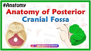
Fav channel for head anatomy till now
Tysm
Thank you so much Ma'am🙂🙂. This video is really helpful for medical students.
Thank you so much mam... really helpful 🙌 Great and clear explanation ❤💯
My pleasure 😊
Best teacher❤❤❤❤❤
God bless you guys
So helpful.Thanks !
you saved my life!!!
Thank you very much... Appreciated...👍👍👍
Amazing 😍
Best teacher 🔥forever
Very well and patiently explained lecs.....Gud job👏
Thanks a lot 😊
Love it
Really thanku mam❤️
Really really very awesome explanation load and loads of thanku ma’am.
Most welcome 😊
Thank u so much ma'am ❣️❣️
Mashallah well explained 😌 very clear concept help me a lot😍
Thanks for liking
Great
Only this time I come to get these things ✨❤️💝🌸🥳
Best
Thankyou
Thank you for the great explanation
You are welcome!
Much much thanks 😊 awesome lecture 👏
Ur most welcome
Thank u mam💕
Thanks mam
Maam may Allah bless you and your family.. thank you maam..
Thank you too
Very nice MAM 👍💐
It's my pleasure
May Allah bless you.
Amazing!!!!
Thank you! Cheers!
Amazing presentation
Thanks a lot
Very well explained... Easy to understand 👍👍👍
Glad to hear that
R u a dental student
Best video 👍🙏
Thanks a lot
Thank you so much 💗
You are so welcome
Tq mam❤️
Thank you very much mam
Most welcome 😊
Nice one sir
Thanks
Good explain
Thanks and welcome
Very nice explanation mam
It's my pleasure
Thankyou mam . We can study clearly without seeing it directly in the lab 🙏🙏
I guess Im asking randomly but does someone know of a way to log back into an Instagram account??
I was stupid forgot my password. I appreciate any help you can give me
@Kingston Douglas instablaster ;)
@Omar Tadeo thanks for your reply. I found the site thru google and I'm waiting for the hacking stuff now.
Seems to take quite some time so I will reply here later with my results.
@Omar Tadeo it worked and I now got access to my account again. I'm so happy:D
Thanks so much, you saved my account :D
@Kingston Douglas no problem =)
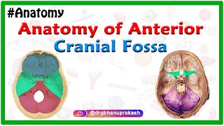
inferior opthalmic vein passes through inf orbital fissure?
😍😍
Mam would you plz tell which sources have you used to compile this lecture....
bd churasia
❤❤❤❤❤❤
😮
Best
Thank you so much mam
Most welcome 😊