Optical Coherence Tomography - OCT | part-1

Вставка
- Опубліковано 31 жов 2019
- INTRODUCTION:
-------------------------
• OCT is an optical instrument that can perform cross-sectional image of biological tissue within less than 10 micron axial resolution using light waves
• Retina is easily accessible to the external light, hence it is specially suited for retinal disorder
• The information provided by OCT is similar to in vivo histopathology of the retina
EXAMPLE:
Zeiss stratus OCT
Topcon 3D OCT-1000
PRINCIPLE OF OCT:
--------------------------------
• It is a imaging technology projected light beam (820nm) near infrared light
• The beam is then split into two beam (Probe beam & Reference beam) by Beam splitter
• Probe beam reach to the target tissue (retina) & reference beam reach to the reference mirror
at a known distance
• The echo time delay of light reflected various layer of target tissue (retina) is compared with
the echo time delay of light reflected from the reference mirror
• A positive interference is produced when light reflected from target tissue & reference mirror
arrives simultaneously
• This interference is measured by a photodetector which finally produce a range of time delays
for comparison
• The interferometer integrates several data points over 2mm depth to construct a tomogram
of retinal structures
• It is real time tomogram using false color scale & different colors represent light
backscattering from the different layers of retina
OCT SYSTEM CONSIST OF:
------------------------------------------
• Fundus viewing unit
• Interferometer unit
• Computer display
• Control panel
• Color inkjet printer
GENERATION OF
----------------------------
• OCT- 1:
o 1st generation
o Transverse resolution 20 micron
o Axial resolution 10 micron
• OCT-2:
o 2nd generation
o Transverse resolution 20 micron
o Axial resolution 10 micron
o Better user interference
• BOTH OCT-1 & OCT-2:
o acquire 100 vertical scan in approximately 1.2 sec
• OCT-3:
o 3rd generation
o Axial resolution 7-8 micron
o Acquire 512 vertical scan
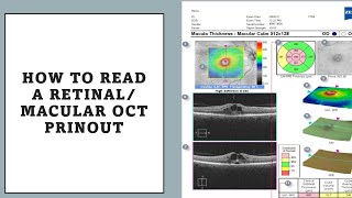
COLOR CODING OF OCT SCAN:
------------------------------------------------
• RED-YELLOW COLORS: represents areas of maximal optical reflection & backscattering
• BLUE & BLACK: Represents areas of minimal optical refelection & backscattering
VARIOUS PATTERN OF B-SCAN:
--------------------------------------------------
• CIRCULAR SCAN FOR THE ONH RNFL:
This generates a plot of the peripapillary RNFL thickness which is important in
glaucoma diagnosis & monitoring
• RADIAL LINE THROUGH ONH:
Consist of 6-24 slices through a common central point on the ONH
• MACULAR RADIAL LINES: Used to measure retinal thickness
FOR DETAILS NOTE;
VISIT OUR WEBSITE: www.smartopto.blogspot.com
OUR OTHERS VIDEOS:
Binocular Subjective Refraction:
Part-1: • Binocular Subjective r...
Part-2: • Septum Method - Turvil...
Part -3: • Polarization Method - ...
Part -4: • Fogging Method - Binoc...
Part-5: • Steps of Binocular Sub...
Biometry:
Part-1: • Introduction Of Biomet...
Paet-2: • Biometry-2 | Measureme...
Part-3 • Biometry-3 | Measureme...
Optical Coherence Topography:
Full: • Optical Coherence Tomo...
Part-1: • Optical Coherence Tomo...
Part-2: • Optical Coherence Tomo...
Dry Eye:
Part-1: • Dry Eye - Introduction...
Part -2: • Dry Eye - Classificati...
Part -3: • Dry Eye - Clinical Fea...
Part -4: • Dry Eye - Treatment (w...
Part -5: • Tear Film Break-up Tim...
Part- 6: • Schirmer test - Dry Ey...
Full: • Dry Eye - Full Tutorial
Myopia
Part-1: • Myopia - Introduction ...
Part-2: / dvrkegdkxq
Part-3: • Myopia - Clinical Pict...
Part -4: • Myopia - Ocular examin...
Part -5: • Myopia - Management or...


How did you make your video like this? As in what software did you use? I love it and I have an assignment due that would work so well in this format. Thanks for the great video!
Do the OCT scan values vary from session to session? For example, if I get my eyes scanned for OCT scan, 3 times so will the values in it vary from each other? By values I mean RNFL thinkness, disc area. Etc
If all parameters (patient position, canthus height, etc) are kept same changes should not be there. Minimum changes may be there which won't be significant.
Thank you,
Team Smart Optometry.
It is very helpful in these global pendamic thankuuu
It really motivates us.
Thank you,
Team Smart Optometry
Thanks for the interesting presentation. About moment 3:03: is it user "interference" or "interface"?
It's interface..
Thank you for your cooperation..
Team "Smart Optometry"
Thanks . 🙏
Stay with us for more Study Materials.
Thank you,
Team Smart Optometry.
I need an urgent help to read a oct report
What is the full form of RNFL
RNFL is "Retinal Nerve Fibre Layer"
Onh is optic nerve head and not hypoplasia
Thanks for your suggestion...
Can you read my oct report sir I need help.
You are suffering from which eye disabiliteis
@@theateam5453 I am not suffering from any disabilities but when I went for the problem of eye floater in left eye . Doctor told me that you have to do oct test after doing that they said to me that you have earlu glaucoma. I was shocked that i am only 23 how can this be possible? So I went for another doc and he said to me that you don’t have glaucoma but revisit after 20 days.
I didn’t shown the oct report to my second doctor. I will show him that report after 20 days.
@@theateam5453 after oct
@@theateam5453 sir one of my eye doctorn said that you have early glaucoma after doing my oct test. I was shocked that how is it possible I am just 23 year old. I just wanted that if you could read my oct it will be very helpful.
Please upload topography video
Thank you for your suggestions. We will try our best.
Thank you,
Team Smart Optometry.
Sir hindi me isaki video bnaiye
Thank you for your suggestion, we will try to make video in Hindi. Optometry is a English based Course. That's why we are preparing video in English.
Thank you,
Team Smart Optometry.
please make videos urdu language