
- 10
- 181 696
CaseStacks
United States
Приєднався 6 вер 2018
Visit CaseStacks.com for 1,500+ interactive cases covering "must-know" diagnoses. Cases include sample reports, focused discussions, and annotated images.
Approach to Head CT and Brain MRI
Dr. Carol Geer (Neuroradiologist at Wake Forest) provides an approach to head CT and brain MRI! This video is ideal for beginners.
Introduction: 0:00
Generation of a CT scan: 0:37
Tissues/Structures on CT: 2:08
Common Pathology: 8:30
MRI: 33:35
👋🏼 Visit CaseStacks.com for 1,500+ interactive cases, a complete radiology call-prep curriculum, and anatomy resources.
~
🐦Twitter/X: x.com/CaseStacks
📸 Instagram: casestacks
#MedEd #FOAMed #FOAMrad #RadRes #Radiology #Imaging #MRI #Anatomy
Переглядів: 376
Відео


New Courses!
Переглядів 4172 роки тому
We are excited to release a new course on brain tumors! 🔗Course: casestacks.com/neuro/fellowship/brain-tumors-course/ 📄Case List: casestacks.com/.../brain-tumors-course/case-index/ 👉 105 Interactive Cases that Include: ➡️ Differentials ➡️ Clinical Pearls ➡️ Focused Discussion ➡️ Annotated Images 👉Earn up to 10.5 CME ➕ We're also releasing a smaller course on neurodegenerative diseases. Both cou...
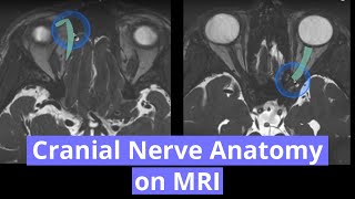
Cranial Nerve Anatomy on MRI
Переглядів 52 тис.3 роки тому
Dr. Tom West (Neuroradiologist at Wake Forest) covers the course of all 12 cranial nerves on MRI! 👇Cranial nerve chapters👇 Introduction: 0:00 1 (Olfactory): 0:12 2 (Optic): 1:03 3 (Oculomotor): 2:38 4 (Trochlear): 4:02 5 (Trigeminal): 6:24 6 (Abducens): 10:14 7 (Facial): 12:42 8 (Vestibulocochlear): 15:44 9 (Glossopharyngeal): 17:17 10 (Vagus): 17:50 11 (Accessory): 18:21 12 (Hypoglossal): 19:3...

Radiology Case Review - A Child Presenting with Dizziness & Headaches
Переглядів 1,3 тис.4 роки тому
This child presented with headache and dizziness. ➡️ Acute cerebellitis is most often postinfectious, occurring 1-2 weeks after a viral infection (e.g. varicella zoster, EBV, measles, HSV, coxsackievirus). ➡️ It most commonly occurs in children and adolescents. ➡️ Neuroimaging is reportedly abnormal in only about 10% of cases where imaging is obtained, so the prevalence of this diagnosi...

Radiology Case Review & Quiz
Переглядів 2,1 тис.4 роки тому
What metabolic disorder classically can cause restricted diffusion in the insula and cingulate gyri?⠀👇Answer below ⠀ 🔗ANSWER: bit.ly/3auoYBv 🔗More MRI cases at CaseStacks.com/neuro/mri/ 🐦Twitter: CaseStacks 📸 Instagram: casestacks #RadRes #MedEd #Radiology #Imaging #Neuroradiology #MRI #FOAMrad #FOAMed #Neurology

Venous Thrombosis on Head CT
Переглядів 20 тис.4 роки тому
Introduction to venous thrombosis on head CT, including: ✔️ Unique challenges & risk factors ✔️ Anatomy and venous drainage territories ✔️ Direct and indirect findings ✔️ Case Examples 👋🏼 Visit CaseStacks.com for 1,500 interactive cases and a complete radiology call-prep curriculum. 🎨 Illustrations by Valerie George, MD. ~ 🐦Twitter: CaseStacks 📸 Instagram: casestacks ...

Hemorrhage on Head CT
Переглядів 2,2 тис.4 роки тому
This short video covers the evolution of the density of intracranial hemorrhage on head CT. 👋 Visit CaseStacks.com for 1,400 interactive cases and a complete call-preparation curriculum. 🐦Twitter: CaseStacks 📸 Instagram: casestacks #RadRes #MedEd #FOAMrad #FOAMed #Neuroradiology #Radiology #Neurosurgery #Neurology

Ischemia on Head CT
Переглядів 5 тис.4 роки тому
This video reviews the findings of early ischemia on head CT, covering: ✔️ Clinical subtypes of ischemia ✔️ Role of non-contrast head CT in stroke workup ✔️ Dense MCA sign ✔️ Pathophysiology of ionic edema ✔️ Blurred basal ganglia sign ✔️ Insular ribbon sign 🔗All cases in this video are available with case write-ups at www.casestacks.com 🎨 Illustrations by Valerie George, MD 🐦Twitter: twitter.c...

Cervical Spine Trauma on CT
Переглядів 11 тис.4 роки тому
Introduction to CT imaging of cervical spine trauma, including: ✔️ Bony and ligamentous anatomy of the atlanto-axial and atlanto-occipital joints ✔️ Atlanto-occipital dislocation ✔️ Occipital condyle, C1 burst, and odontoid fractures. 🔗All cases in this video are available with case write-ups at www.casestacks.com 🎨 Illustrations by Valerie George, MD. 🐦Twitter: CaseStacks 📸 Instagr...
CaseStacks.com | Learn & Practice Radiology
Переглядів 87 тис.4 роки тому
Over 1,400 interactive cases covering "must know" diagnoses. Cases include sample reports, focused discussion & annotated images | 🔗 CaseStacks.com 🐦Twitter: CaseStacks 📸 Instagram: CaseStacks 💌 Sign up for our newsletter at casestacks.com/account/newsletter/ We'd love to hear from you. Direct message us on Twitter @CaseStacks or feel free to email us at user@casestack...
Hi, I have not normal case with CVST if you can help. Thx
someday , someone will develop decent webinar audio quality. I just know it.
Thank you so much 🎉🎉🎉
*get 4 views
Thank you so much
nicely explained
👏🏻👏🏻
very nice, thank you very much. One little correction; the part where the fourth cranial nerve crosses is called the frenulum, the superior medullary velum is more caudal at the level of the superior cerebellar peduncles
Hope you can explain temporal lobe in detail. Maybe
I lost my 15 years son with these diseas 15 days back
I appreciate the concise detailed video. It shows you have great grasp on the subject
Amazing video, thank you!
great content, do you have any dataset with ground truth available for AI applications? Thank you
Chernobyl
Very useful for me, thanks
Great thank you
❤
Treatment of mesial e epelesy is by surgical removal of anciss gyrus
Thank you for extraordinary lecture
Most welcome!
Excellent!
Many thanks!
Great
Thank you for tis video
How to make a PPT that can directly scroll images like in the video? Really need it
لطفا ویدیو رو با زیرنویس بگذارید مرسی
Thank you very good
Thank you sir, very nice Explanation
I did not understand the video, but I came here coz of headache behind my neck region extends way up to top center
Amazing! Thank you!
What power of mri is that 7.0 tesla?
FIESTA sequence
Is there a part 2?
Verry nice .
-what about the primordial stability ligament ' THe Apical ligament ' allows for flexion and extension ' stress tests on dead Vietnam soldiers in the 60s confirmed the main stability ligament .
This is so far the best mir anatomical video I have watched on UA-cam
What I do Iam pains, I can't stand ordering, or walk without a stick, I can't sleep, way out
Great Video and one of the most informative I have ever seen. Many thanks.
I have cerebral artery compression of trigeminal nerve root. It is awful.
What are your symptoms ?? And what is reason for compression doctor told you anything
Symptoms?
superb presentation
Great
most detail radiology anatomy of cranial nerve in youtube I have ever seen.
great
Trick: Arterial thrombus appears black than the artery or lumen of the artery while veinous thrombus appears white than vein as artery appears hyperdense so thrombus can be seen only when only when it is aged i.e. not new while veins being hypodense does appreciate a thrombus when it is new . Please o please correct me IF i' m wrong.
Not really. Hyperdense thrombus in arteries is more dense than surrounding blood. Logically, thrombus surrounded by blood would look the same regardless if arterial or venous
Do i iave more exp.
When you place the cushion between the artery and the nerve, does the cushion ever move around the brain years later and cause any problems?
Yes then again mvd is repeated
I think some Neurosurgeons use some type of sling to separate the blood vessel and the facial nerve. I would think that would be more permanent than Teflon pads.
Re: @3:25 DEEP system "Vein of Galen" drains... deep white matter of Frontal lobes Temporal lobes Pariental lobes Corpus Collostum Upper Brain Stem Basal Ganglia Thalami (?) 1. What are these responsible for? 2. If it drains outwardly, through sinuses, can it receive treatment inwardly, through the same sinuses? If it can go out one path, it can go in through the same path, I assume? Just wondering
Brilliant share Sir.
Todo un reto en la materia de la medicina 🌎💛☕🌻🙏🍓
Please make move videos like this,helpful for us.👍👍👍
Thx Sir I really like your way
A good presentation.