
- 142
- 792 368
theRadiologist
Приєднався 1 сер 2018
Hi, I'm Dr Naveen Sharma and I'm a Consultant Radiologist based in the UK. Before becoming a radiologist I found the topic really daunting and to be honest had no idea what I was looking at when faced with an X-ray or scan on the ward. So in 2018 I started an Instagram account teaching radiology which I tried to make as simple as possible and it seemed to be helpful, now reaching nearly 400k followers.
I started this UA-cam channel to try and go into a bit more depth so as well as short case videos you'll find more detailed guides on how to look at scans and X-rays - basically this is all the stuff that I wish someone had told me when I was learning so hope it comes into use for anyone who is trying to learn some radiology.
I am trying to post something new here each week so if you find it useful, please support me by subscribing and also please do leave a comment to let me know what you would find useful to see on the channel.
Chest X-Ray breakdown: understanding mediastinal contour
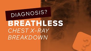
A male in his 30s presents with chest pain. What does this PA chest X-Ray show?
OESOPHAGEAL DILATATION - ACHALASIA
👨🏽💻 The key to this X-Ray is spotting the abnormal right sided mediastinal contour and understanding when you see this involve the whole length of the right side of the mediastinum this could represent oesophageal dilatation. Placing this outside of the middle mediastinum lends weight to this .
👨🏽💻Achalasia is an oesophageal motility disorder characterized by impaired relaxation of the lower esophageal sphincter and absent peristalsis in the esophageal body. Imaging typically reveals dilation of the esophagus with a tapered narrowing at the gastroesophageal junction, giving rise to a classic “bird’s beak” appearance on barium swallow.
🔻@theradiologistpage
----
✅Patient consent obtained
#theradiologist #radiology #radiologist #physician #physicianassistant #medicine #medstudent #medicalstudent #medschool #medicalschool #radtech #xray #medical #chestxray #doctor #medicalstudents #frcr
Переглядів: 13 667
Відео


Chest X-Ray breakdown: assessing a pneumothorax
Переглядів 9 тис.9 місяців тому
A male in his 50s presents with acute breathlessness. What does the X-Ray show? HYDROPNEUMOTHORAX AND RIGHT LUNG OPACITIES 👨🏽💻Another tricky X-Ray! There is a lot going on on this X-Ray and so it shows the importance of a systematic approach. Firstly there is a hydropneumothorax: we can see gas in the pleural space evidenced by a lack of lung markings and pleural line. At the right lung base w...

Chest X-Ray breakdown: assessing multiple lung nodules
Переглядів 10 тис.Рік тому
A female in her 40s presents with chest pain and has an X-Ray. What does it show and what is the differential? BENIGN METASTASISING LEIOMYOMAS 👨🏽💻A difficult diagnosis - this case is all about recognising the lung nodules and coming up with a sensible differential and management plan. Lung metastases need to be considered and in a patient of this age, breast cancer is a possible primary 👨🏽💻Th...

CT head breakdown: a guide to subarachnoid haemorrhage
Переглядів 4,4 тис.Рік тому
A female in her 50s presents after a fall and loss of consciousness and has a CT head. What does it show? SUBARACHNOID HAEMORRHAGE 👨🏽💻This CT is about first spotting the subarachnoid haemorrhage - look for bright density material occupying the basal cisterns and sulci. Particularly look at the pentagon shaped suprasellar cistern 👨🏽💻Then look for the cause - most patients go on to have a CT an...

Chest X-Ray breakdown: looking for difficult lung nodules
Переглядів 8 тис.Рік тому
A female smoker in her 50s presents with a cough and has a chest X-Ray. What does it show? OBSCURED LUNG NODULE 👨🏽💻This X-Ray is all about spotting nodules in tricky areas. If the nodule had been slightly higher or lower this would have been an easy spot but given it was behind the clavicle means it was a lot more tricky 👨🏽💻 Some review areas on chest X-Ray need you to assess the central stru...

CTPA breakdown: when the filling defects don’t represent a PE
Переглядів 5 тис.Рік тому
A male in his 70s presents to the Emergency Department with worsening breathlessness and has a CTPA. What does it show? PULMONARY ARTERY SARCOMA 👨🏽💻This is a difficult diagnosis, one that we do not see too often but something that is worth bearing in mind when the clinical picture doesn’t quite fit a PE and filling defects within the pulmonary arteries persist 👨🏽💻If you see a PE on CTPA there...

Abdominal CT breakdown: explaining the sandwich sign
Переглядів 6 тис.Рік тому
Abdominal CT breakdown: explaining the sandwich sign
Chest X-Ray breakdown: a guide to pulmonary oedema
Переглядів 8 тис.Рік тому
Chest X-Ray breakdown: a guide to pulmonary oedema

Chest X-Ray breakdown: a guide to lung adenocarcinoma
Переглядів 8 тис.Рік тому
Chest X-Ray breakdown: a guide to lung adenocarcinoma
Chest X-Ray breakdown: How to assess the posterior mediastinum on Chest X-Ray
Переглядів 12 тис.Рік тому
Chest X-Ray breakdown: How to assess the posterior mediastinum on Chest X-Ray
MRI Head breakdown: looking for a cause of hydrocephalus
Переглядів 3,9 тис.Рік тому
MRI Head breakdown: looking for a cause of hydrocephalus
CTPA breakdown: looking for other causes of breathlessness
Переглядів 7 тис.Рік тому
CTPA breakdown: looking for other causes of breathlessness
Chest X-Ray breakdown: looking for subtle abnormalities
Переглядів 8 тис.Рік тому
Chest X-Ray breakdown: looking for subtle abnormalities
Chest X-Ray guide: how to assess the hilar regions
Переглядів 7 тис.Рік тому
Chest X-Ray guide: how to assess the hilar regions
CT head breakdown: assessing high density lesions
Переглядів 6 тис.Рік тому
CT head breakdown: assessing high density lesions
Chest X-Ray breakdown: assessing lung nodules in young patients
Переглядів 8 тис.Рік тому
Chest X-Ray breakdown: assessing lung nodules in young patients
CT Head breakdown: how to assess for acute stroke on CT
Переглядів 6 тис.Рік тому
CT Head breakdown: how to assess for acute stroke on CT
Chest X-Ray breakdown: assessing the lung apices
Переглядів 7 тис.Рік тому
Chest X-Ray breakdown: assessing the lung apices
Abdominal CT breakdown: assessing back pain
Переглядів 6 тис.Рік тому
Abdominal CT breakdown: assessing back pain
Chest X-Ray breakdown: clearing the retrocardiac region
Переглядів 6 тис.Рік тому
Chest X-Ray breakdown: clearing the retrocardiac region
Abdominal CT breakdown: assessing small bowel tumours
Переглядів 5 тис.Рік тому
Abdominal CT breakdown: assessing small bowel tumours
Finger pain post hyperextension: how to assess volar plate injuries on X-Ray
Переглядів 3 тис.Рік тому
Finger pain post hyperextension: how to assess volar plate injuries on X-Ray
Chest X-Ray breakdown: assessing pericardial calcification
Переглядів 6 тис.Рік тому
Chest X-Ray breakdown: assessing pericardial calcification
Chest X-Ray breakdown: assessing the azygos contour
Переглядів 6 тис.Рік тому
Chest X-Ray breakdown: assessing the azygos contour
Chest X-Ray breakdown: assessing a film with pleural plaques
Переглядів 13 тис.Рік тому
Chest X-Ray breakdown: assessing a film with pleural plaques
Abdominal X-Ray breakdown: a guide to volvulus on imaging
Переглядів 4,4 тис.Рік тому
Abdominal X-Ray breakdown: a guide to volvulus on imaging
Chest X-Ray breakdown: a guide to bronchiectasis on imaging
Переглядів 12 тис.Рік тому
Chest X-Ray breakdown: a guide to bronchiectasis on imaging
Chest X-Ray breakdown: assessing external objects
Переглядів 4,7 тис.Рік тому
Chest X-Ray breakdown: assessing external objects
Chest X-Ray breakdown: How to approach the hilar regions
Переглядів 6 тис.Рік тому
Chest X-Ray breakdown: How to approach the hilar regions
Ankle X-Ray breakdown: How to approach a bone lesion on X-Ray
Переглядів 6 тис.Рік тому
Ankle X-Ray breakdown: How to approach a bone lesion on X-Ray
2024
😂😂
true
thank you
very helpful thank u theradiologist
Highly underrated channel. Please make a video on Abdominal CT approach and brain MRI approach
😅like
Everybody in here report at 9 am in the morning
You’re awesome
Thx, this was extremely helpful. Isn't it better to look for the gastric bubble if there is no label for ap or pa?
Ливер? так на видео живот, интересно
❤❤❤
I know the feeling. I did that until I tried radiology, and everything just clicked! I am now in my 3rd year of Radiology. I have 2 and a half years left to become a specialist.
Now these are what I call Kawaii (beautiful) lessons!! A lot of info in 2 mins, connecting all the dots... Amazing!!
😂😂😅
Hi, I would like to know your opinion about my case if possible
Japan entered the chat
does it cause by smoking
I’m becoming a radiology therapist in the future!!
😂😂😂😂😂
this video was helpful!
This is an extremely well done video!
I want more videos like thiz
thanks very much!
Thank you for doing this video! It is excellent!
At least it’s better than sitting in a dark room reading images but doing nothing much else😂
at least it is much better than dying from radiation 😢
Excellent video! Very helpful!
Thanks for all doctor ❤ Can you plz tell me what app do you use to create these videos ?
Thank you for useful lecture. Do the dots I see at 0:21 just mean nothing? Are they just defects? I see them a lot and sometimes I get confused ;
Bilateral lower lobe pneumonia possibly with a small rt-sided abscess
Y’all can be real dramatic with these hide-n-seek games 😂😂😂.
Ryt on point 😂😂😂😂😂😂
Wow...our bodies work in mysterious ways
can u tell me why is it hard?
Proud
Nice one !
Sounds similar.
Very nice ❤
Tq
Great case
Thanks for watching!
ever heard of a Lateral CXR...???
Yes 😂 In some places in the world laterals are not routinely done, like the UK. Not with any good reason it should be said. So a lot of my content focuses on how to get the best out of a frontal film. In this case a dilated oesophagus has a typical appearance on a frontal X-Ray so I really wanted to highlight that
You make me love Radiology ❤
So so good
Educative!
Is the trachea shift to right side?? Am i right or wrong?
You are right. If you look at the coronal slices of the CT scan on the video you’ll see superiorly the oesophagus is very dilated and quite central pushing the trachea to the right before the oesophagus takes a position on the right side of the mediastinum more inferiorly
What makes the esophagus shift lateral to the right?
Pneumothorax, Fibrosis
It’s a good question, I’ve only ever seen it move to the right. There may be a more accomplished answer but I think there are too many structures on the left for it to move that side once it becomes dilated
Why not hiatal hernia?
Good thought although I’ve never seen a hiatus hernia extend up to the clavicle
👌 Well explained
Thanks for watching!
Amazing work! Please keep on doing them! Thank you so much for helping us!
Good case and very nicely explained 👍